A collection of videos relating to the diagnosis and treatment of eye movement disorders. This collection includes many demonstrations of examination techniques.
Dan Gold, D.O., Associate Professor of Neurology, Ophthalmology, Neurosurgery, Otolaryngology - Head & Neck Surgery, Emergency Medicine, and Medicine, The Johns Hopkins School of Medicine.
A collection of videos relating to the diagnosis and treatment of eye movement disorders.
NOVEL: https://novel.utah.edu/
TO
| Title | Description | Type | ||
|---|---|---|---|---|
| 151 |
 |
The Apogeotropic Variant of Horizontal Canal BPPV | 𝗢𝗿𝗶𝗴𝗶𝗻𝗮𝗹 𝗗𝗲𝘀𝗰𝗿𝗶𝗽𝘁𝗶𝗼𝗻: This is a patient with the apogeotropic (nystagmus beating towards the sky) variant of right horizontal canal (HC) benign paroxysmal positional vertigo (BPPV). In a patient with geotropic (nystagmus beating towards the gr... | Image/MovingImage |
| 152 |
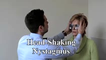 |
Head-Shaking Nystagmus | Head-shaking nystagmus: With a peripheral lesion, similar to vibration, transiently accentuates vestibular asymmetry when baseline VOR function is asymmetric, central patterns are well described and have localizing value (e.g., causing vertical nystagmus after horizontal head-shaking, horizontal nys... | Image/MovingImage |
| 153 |
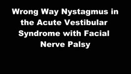 |
Wrong Way Nystagmus - Cranial Nerve 7 and 8 | Image/MovingImage | |
| 154 |
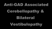 |
Anti-GAD Associated Cerebellopathy and Bilateral Vestibulopathy | This is a 70-year-old woman with the subacute onset of severe imbalance and dizziness. On her initial examination, she had prominent gaze-evoked nystagmus and bilateral vestibular loss. Smooth pursuit was saccadic, although her vestibulo-ocular reflex (VOR) suppression was much smoother. Usually pur... | Image/MovingImage |
| 155 |
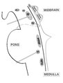 |
Brainstem Ocular Motor Machinery | Seen here is a sagittal view of the brainstem. The medulla has a significant role in gaze-holding, and the nucleus prepositus hypoglossi (NPH, along with the medial vestibular nucleus ) is the horizontal neural integrator. The abducens (6th) nucleus is located in the dorsal pons, and sends off the 6... | Image/MovingImage |
| 156 |
 |
Trigeminal Motor Neuropathy with Weakness and Atrophy of the Muscles of Mastication | This is a man who was diagnosed with polio in childhood, which involved the motor (VIII) division of the right trigeminal nerve. The motor portion of the trigeminal nerve innervates the muscles of mastication (temporalis, masseter - both of which demonstrate wasting in this patient - as well as the ... | Image/MovingImage |
| 157 |
 |
Periodic Alternating Nystagmus Due to Spinocerebellar Ataxia Type 6 | 𝗢𝗿𝗶𝗴𝗶𝗻𝗮𝗹 𝗗𝗲𝘀𝗰𝗿𝗶𝗽𝘁𝗶𝗼𝗻: This 50-yo-man complained of imbalance for several years and more recently oscillopsia. On examination, there was saccadic pursuit and VOR suppression in addition to gaze-evoked nystagmus with rebound, raising suspicion f... | Image/MovingImage |
| 158 |
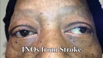 |
INOs in Stroke | 𝗢𝗿𝗶𝗴𝗶𝗻𝗮𝗹 𝗗𝗲𝘀𝗰𝗿𝗶𝗽𝘁𝗶𝗼𝗻: This video shows 3 patients with vascular risk factors who suffered strokes of the MLF resulting in unilateral INO in each case. In the second case, INO was diagnosed status post cardiac catherization and MRI was found to... | Image/MovingImage |
| 159 |
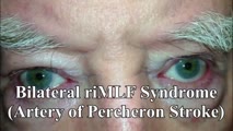 |
riMLF Syndrome from Artery of Percheron Stroke | 𝗢𝗿𝗶𝗴𝗶𝗻𝗮𝗹 𝗗𝗲𝘀𝗰𝗿𝗶𝗽𝘁𝗶𝗼𝗻: This is a 65-yo-man who suffered the abrupt onset of loss of consciousness followed by difficulty looking down. MRI showed bilateral rostral midbrain strokes in the distribution of the artery of Percheron. He could not in... | Image/MovingImage |
| 160 |
 |
Unilateral 3rd, 4th, and 6th Nerve Palsies Due to Cavernous Sinus Meningioma | 𝗢𝗿𝗶𝗴𝗶𝗻𝗮𝗹 𝗗𝗲𝘀𝗰𝗿𝗶𝗽𝘁𝗶𝗼𝗻: This is a 50-year-old woman presenting with a partial 3rd nerve palsy (mild pupil involvement), partial 6th nerve palsy, and no clear incyclotorsion with downgaze, suggestive of additional 4th nerve palsy, all on the left... | Image/MovingImage |
| 161 |
 |
Central 4th Nerve Palsy with Contralateral Horner's Syndrome | This is a 60-yo-woman who presented with a complaint of diplopia. Examination demonstrated a left hypertropia that worsened in right and down gaze as well as in left head tilt, and a left 4th nerve palsy was diagnosed. There was also evidence of a mild motility deficit in down/medial gaze OS, consis... | Image/MovingImage |
| 162 |
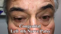 |
Inferior Oblique Overaction in a Congenital 4th Nerve Palsy | 60-yo-man complaining of intermittent oblique diplopia. There was a left hypertropia that worsened in down gaze, right gaze and in left head tilt. There was a large vertical fusional amplitude in addition to a longstanding rightward head tilt, and on examination there was left inferior oblique overa... | Image/MovingImage |
| 163 |
 |
Complete Microvascular 6th Nerve Palsy with Slow Abducting Saccade | 𝗢𝗿𝗶𝗴𝗶𝗻𝗮𝗹 𝗗𝗲𝘀𝗰𝗿𝗶𝗽𝘁𝗶𝗼𝗻: This is a 90-year-man with HTN, HLD, DM who woke up with horizontal diplopia. Two years prior, he was diagnosed with a microvascular right 6th nerve palsy that resolved over several months. There was little concern for gi... | Image/MovingImage |
| 164 |
 |
One-and-a-Half Syndrome Due to Pontine Hemorrhage | This is a 50-year-old woman who, while exercising in the gym, suddenly experienced vertigo, nausea, vomiting, tingling in the left arm, and diplopia. MRI demonstrated a brainstem hemorrhage that involved the right greater than left pons. Examination demonstrated a right horizontal gaze palsy due to ... | Image/MovingImage |
| 165 |
 |
Leukemic Leptomeningeal Carcinomatosis Causing 4th and 6th Nerve Palsies | 𝗢𝗿𝗶𝗴𝗶𝗻𝗮𝗹 𝗗𝗲𝘀𝗰𝗿𝗶𝗽𝘁𝗶𝗼𝗻: This is a 55-yo-man with CML that recurred as AML. Diagonal diplopia developed, and on examination he was found to have a partial right 6th nerve palsy, in addition to a left hypertropia that increased in right gaze, down... | Image/MovingImage |
| 166 |
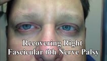 |
Slow Abducting Saccade in 6th Nerve Palsy | 40-yo-man with a right fascicular 6th nerve palsy due to stroke. There was improvement and only a minimal residual right abduction paresis OD by this visit, but still a relatively slow right abducting saccade seen in the video, especially apparent in the slow motion segment. Video shows slow abduct... | Image/MovingImage |
| 167 |
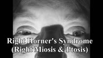 |
Horner's Syndrome with Anhidrosis | 𝗢𝗿𝗶𝗴𝗶𝗻𝗮𝗹 𝗗𝗲𝘀𝗰𝗿𝗶𝗽𝘁𝗶𝗼𝗻: This is a patient with the onset of ptosis OD years prior, with clear evidence of a Horner's syndrome. Imaging of the oculosympathetic tract was unrevealing. The patient also mentioned that with exercise, the left side of... | Image/MovingImage |
| 168 |
 |
Apraclonidine Testing in Horner's syndrome | This patient experienced relatively abrupt ptosis and was seen and diagnosed with a Horner's syndrome within a few days of the onset. There were no other exam findings and history did not offer clues as to the etiology. Neuroimaging of the oculosympathetic tract was unrevealing. Apraclonidine testin... | Image/MovingImage |
| 169 |
 |
Skew Deviation and the Triad of the Ocular Tilt Reaction (OTR) | This is a patient who presented with vertical diplopia, who was found to have a complete ocular tilt reaction including the following features: (1) Skew deviation - right hypertropia that was about 30 prism diopters in all directions of gaze including right, left, up, down, as well as in right and ... | Image/MovingImage |
| 170 |
 |
Mesodiencephalic Stroke Causing Unilateral riMLF and INC Ocular Motor Syndromes | 𝗢𝗿𝗶𝗴𝗶𝗻𝗮𝗹 𝗗𝗲𝘀𝗰𝗿𝗶𝗽𝘁𝗶𝗼𝗻: This is a 65-year-old man who experienced the sudden onset of diplopia (with horizontal and vertical components), dysarthria and imbalance. An MRI performed the following day showed a left mesodiencephalic stroke. The pat... | Image/MovingImage |
| 171 |
 |
Medial Longitudinal Fasciculus Syndrome with Prominent Spontaneous Nystagmus | 𝗢𝗿𝗶𝗴𝗶𝗻𝗮𝗹 𝗗𝗲𝘀𝗰𝗿𝗶𝗽𝘁𝗶𝗼𝗻: This is a 60-year-old man who experienced the abrupt onset of diplopia and imbalance. He had typical features of a left medial longitudinal fasciculus (MLF) syndrome including left internuclear ophthalmoplegia (INO) and l... | Image/MovingImage |
| 172 |
 |
Paroxysmal Ocular Tilt Reaction | 𝗢𝗿𝗶𝗴𝗶𝗻𝗮𝗹 𝗗𝗲𝘀𝗰𝗿𝗶𝗽𝘁𝗶𝗼𝗻: This is a 60-year-old woman who 2 years prior experienced a left sided hypertensive hemorrhagic stroke, resulting in right hemiparesis, dysarthria and vertical diplopia. The initial vertical diplopia resolved completely a... | Image/MovingImage |
| 173 |
 |
Alternating Hypertropias - Bilateral 4th Nerve Palsies and Alternating Skew Deviation | 𝗢𝗿𝗶𝗴𝗶𝗻𝗮𝗹 𝗗𝗲𝘀𝗰𝗿𝗶𝗽𝘁𝗶𝗼𝗻: Seen here are two patients with alternating hypertropias. The first is a 70-year-old woman with a diagnosis of cerebellar ataxia, neuropathy, vestibular areflexia syndrome (CANVAS). In the video, both spontaneous downbeat... | Image/MovingImage |
| 174 |
 |
Isolated Central 4th Nerve Palsy | 𝗢𝗿𝗶𝗴𝗶𝗻𝗮𝗹 𝗗𝗲𝘀𝗰𝗿𝗶𝗽𝘁𝗶𝗼𝗻: This is a 40-year-old man with a right hypertropia that worsened in left and down gaze in addition to right head tilt, and improved in left head tilt. There was subjective excyclotorsion OD with double Maddox rod testing.... | Image/MovingImage |
| 175 |
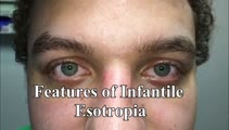 |
Eye Signs in Infantile Esotropia - Latent Nystagmus and Inferior Oblique Overaction | 𝗢𝗿𝗶𝗴𝗶𝗻𝗮𝗹 𝗗𝗲𝘀𝗰𝗿𝗶𝗽𝘁𝗶𝗼𝗻: This is a 25-yo-man with a history of amblyopia and intermittent eye crossing. On exam, he had a comitant 25 prism diopter esotropia, and other features of infantile (or congenital) esotropia including: latent nystagmus (... | Image/MovingImage |