A collection of videos relating to the diagnosis and treatment of eye movement disorders. This collection includes many demonstrations of examination techniques.
Dan Gold, D.O., Associate Professor of Neurology, Ophthalmology, Neurosurgery, Otolaryngology - Head & Neck Surgery, Emergency Medicine, and Medicine, The Johns Hopkins School of Medicine.
A collection of videos relating to the diagnosis and treatment of eye movement disorders.
NOVEL: https://novel.utah.edu/
TO
Filters: Collection: "ehsl_novel_gold"
| Title | Description | Type | ||
|---|---|---|---|---|
| 151 |
 |
Abnormal Visually-Enhanced VOR in Cerebellar Ataxia, Neuropathy, Vestibular Areflexia Syndrome (CANVAS) | A 67 year old woman presented with 1 year of progressive numbness, gait instability, and oscillopsia when walking or with head movements. Examination showed excessive square-wave jerks, bilateral horizontal gaze-evoked nystagmus, impairment of the visually-enhanced vestibular ocular reflex (vVOR - s... | Image/MovingImage |
| 152 |
 |
Nystagmus Due to Paraneoplastic (Anti-Yo) Brainstem and Cerebellar Degeneration | 𝗢𝗿𝗶𝗴𝗶𝗻𝗮𝗹 𝗗𝗲𝘀𝗰𝗿𝗶𝗽𝘁𝗶𝗼𝗻: This is a 40-yo-woman with anti-Yo antibody associated with ovarian cancer. Initial symptoms 2.5 years prior (to this video) included imbalance and dysarthria. She complained of oscillopsia which was due to her upbeat nys... | Image/MovingImage |
| 153 |
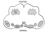 |
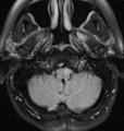
Medullary Structures Relevant to the Ocular Motor and Vestibular Consequences of Lateral Medullary (Wallenberg) Syndrome | This is an axial section of the medulla showing the structures that, when damaged, are responsible for the vestibular and ocular motor features of the lateral medullary or Wallenberg syndrome. The nucleus prepositus hypoglossi (NPH) and medial vestibular nucleus (MVN) complex is important for horizo... | Image |
| 154 |
 |
Saccadic Pathways in the Brainstem and Cerebellum & Mechanism for Saccadic Dysmetria in Wallenberg Syndrome - Abnormal Function of the Brainstem/Cerebellar Saccadic Pathways with a Left Wallenberg Syndrome | The end result of a lesion involving the climbing fibers within the left lateral medulla is deficient rightward saccades (contralesional hypometric saccades), and over-active leftward saccades (ipsilesional hypermetric saccades), and ipsilesional ocular lateropulsion given this baseline imbalance. M... | Image |
| 155 |
 |
Ocular Bobbing Due to Hepatic Encephalopathy | 𝗢𝗿𝗶𝗴𝗶𝗻𝗮𝗹 𝗗𝗲𝘀𝗰𝗿𝗶𝗽𝘁𝗶𝗼𝗻: This is a 55-year-old man presented with hepatic encephalopathy, and found to have ocular bobbing. Head CT did not show any acute changes. Ocular bobbing almost always localizes to the pons, although cerebellar pathology ... | Image/MovingImage |
| 156 |
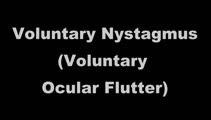 |
Voluntary Ocular Flutter | 𝗢𝗿𝗶𝗴𝗶𝗻𝗮𝗹 𝗗𝗲𝘀𝗰𝗿𝗶𝗽𝘁𝗶𝗼𝗻: This is a 45-yo-man with intermittent complaints of horizontal oscillopsia for 1 year. On examination, all classes of eye movements were normal, and neurologic examination was normal. MRI of the brain had been performed p... | Image/MovingImage |
| 157 |
 |
Ocular Motor & Vestibular Features of the MLF Syndrome | 𝗢𝗿𝗶𝗴𝗶𝗻𝗮𝗹 𝗗𝗲𝘀𝗰𝗿𝗶𝗽𝘁𝗶𝗼𝗻: This 61-year-old woman with HTN and DM presented for evaluation of acute onset diagonal diplopia. Adduction OS was about 60% of normal while medialization OS improved with convergence. In right gaze, dissociated abducti... | Image/MovingImage |
| 158 |
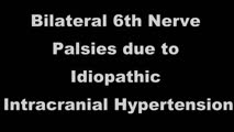 |
Bilateral 6th Nerve Palsies Due to Idiopathic Intracranial Hypertension | This is a 25-year-old woman who presented with diplopia and blurry vision. On exam, she was found to have papilledema and bilateral 6th nerve palsies. Her opening pressure was >40 cm of water with a normal CSF analysis, and neuroimaging was unremarkable aside from subtle findings that have been asso... | Image/MovingImage |
| 159 |
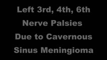 |
Unilateral 3rd, 4th, and 6th Nerve Palsies Due to Cavernous Sinus Meningioma | 𝗢𝗿𝗶𝗴𝗶𝗻𝗮𝗹 𝗗𝗲𝘀𝗰𝗿𝗶𝗽𝘁𝗶𝗼𝗻: This is a 50-year-old woman presenting with a partial 3rd nerve palsy (mild pupil involvement), partial 6th nerve palsy, and no clear incyclotorsion with downgaze, suggestive of additional 4th nerve palsy, all on the left... | Image/MovingImage |
| 160 |
 |
One-and-a-Half Syndrome Due to Pontine Hemorrhage | This is a 50-year-old woman who, while exercising in the gym, suddenly experienced vertigo, nausea, vomiting, tingling in the left arm, and diplopia. MRI demonstrated a brainstem hemorrhage that involved the right greater than left pons. Examination demonstrated a right horizontal gaze palsy due to ... | Image/MovingImage |
| 161 |
 |
Leukemic Leptomeningeal Carcinomatosis Causing 4th and 6th Nerve Palsies | 𝗢𝗿𝗶𝗴𝗶𝗻𝗮𝗹 𝗗𝗲𝘀𝗰𝗿𝗶𝗽𝘁𝗶𝗼𝗻: This is a 55-yo-man with CML that recurred as AML. Diagonal diplopia developed, and on examination he was found to have a partial right 6th nerve palsy, in addition to a left hypertropia that increased in right gaze, down... | Image/MovingImage |
| 162 |
 |
Typical Features of Duane Syndrome Type 1 | 𝗢𝗿𝗶𝗴𝗶𝗻𝗮𝗹 𝗗𝗲𝘀𝗰𝗿𝗶𝗽𝘁𝗶𝗼𝗻: This is a patient seen for vestibular complaints, who on exam, was found to have (unrelated to her vestibular symptoms) impaired abduction OS. In adduction, there was narrowing of the palpebral fissure OS, a result of glo... | Image/MovingImage |
| 163 |
 |
Superior Canal Dehiscence | 𝗢𝗿𝗶𝗴𝗶𝗻𝗮𝗹 𝗗𝗲𝘀𝗰𝗿𝗶𝗽𝘁𝗶𝗼𝗻: This is a 60-yo-man who complained of autophony (eg, hearing his own heartbeat, noting that his own voice sounded too loud) and dizziness triggered with loud noises and straining at times. With pinched-nose Valsalva maneu... | Image/MovingImage |
| 164 |
 |
Vibration and Hyperventilation-induced Nystagmus from Vestibular Schwannoma | 𝗢𝗿𝗶𝗴𝗶𝗻𝗮𝗹 𝗗𝗲𝘀𝗰𝗿𝗶𝗽𝘁𝗶𝗼𝗻: This is a 50-yo-woman with imbalance, and with fixation removed on her examination (with Frenzel goggles), there was no spontaneous nystagmus. Using a handheld vibrator to vibrate the mastoids and vertex, there was a righ... | Image/MovingImage |
| 165 |
 |
Demonstration of HINTS Examination in a Normal Subject | In the acute vestibular syndrome - consisting of acute prolonged vertigo, spontaneous nystagmus, imbalance, nausea/vomiting, head motion intolerance which is typically due to vestibular neuritis or posterior fossa stroke - a 3 step test of ocular motor and vestibular function known as HINTS, has hig... | Image/MovingImage |
| 166 |
 |
Neuro-Ophthalmic Features and Pseudo-MG Lid Signs in Miller Fisher Syndrome | 𝗢𝗿𝗶𝗴𝗶𝗻𝗮𝗹 𝗗𝗲𝘀𝗰𝗿𝗶𝗽𝘁𝗶𝗼𝗻: This is a 51-year-old woman who presented with imbalance, acute onset dizziness and diplopia that developed over three days following two weeks of upper respiratory infection and bacterial conjunctivitis. When she was ini... | Image/MovingImage |
| 167 |
 |
Neuro-Ophthalmic Features and Pseudo-MG Lid Signs in Miller Fisher Syndrome (Figure 1) | This is a 51-year-old woman who presented with imbalance, acute onset dizziness and diplopia that developed over three days following two weeks of upper respiratory infection and bacterial conjunctivitis. When she was initially seen as an outpatient, nystagmus was noted to the right and left, and a ... | Image |
| 168 |
 |
Subtle Torsional Pendular Nystagmus in Oculopalatal Tremor (OPT) | 𝗢𝗿𝗶𝗴𝗶𝗻𝗮𝗹 𝗗𝗲𝘀𝗰𝗿𝗶𝗽𝘁𝗶𝗼𝗻: This is a 50-year-old woman who presented with imbalance, and MRI demonstrated a right cerebellar cavernous malformation. She underwent surgery to resect the malformation, and post-operatively experienced right hemiparesi... | Image/MovingImage |
| 169 |
 |
Subtle Torsional Pendular Nystagmus in Oculopalatal Tremor (OPT) (Figure 1) | This is a 50-year-old woman who presented with imbalance, and MRI demonstrated a right cerebellar cavernous malformation. She underwent surgery to resect the malformation, and post-operatively experienced right hemiparesis and ataxia. Six months after the surgery, balance worsened and vision became ... | Image |
| 170 |
 |
Relationship Between Semicircular Canals and Extraocular Muscles | Figure 1: When stimulated, each of the 6 angular acceleration detecting semicircular canals (3 on the right and 3 on the left) responds with a conjugate eye movement, with the vector(s) indicated below. PC=posterior canal; HC=horizontal (also known as lateral) canal; AC=anterior (also known as super... | Image |
| 171 |
 |
The Geotropic Variant of Horizontal Canal BPPV | 𝗢𝗿𝗶𝗴𝗶𝗻𝗮𝗹 𝗗𝗲𝘀𝗰𝗿𝗶𝗽𝘁𝗶𝗼𝗻: This is a patient with the geotropic (nystagmus beating towards the ground) variant of left horizontal canal (HC) benign paroxysmal positional vertigo (BPPV). In a patient with geotropic (nystagmus beating towards the gro... | Image/MovingImage |
| 172 |
 |
The Apogeotropic Variant of Horizontal Canal BPPV | 𝗢𝗿𝗶𝗴𝗶𝗻𝗮𝗹 𝗗𝗲𝘀𝗰𝗿𝗶𝗽𝘁𝗶𝗼𝗻: This is a patient with the apogeotropic (nystagmus beating towards the sky) variant of right horizontal canal (HC) benign paroxysmal positional vertigo (BPPV). In a patient with geotropic (nystagmus beating towards the gr... | Image/MovingImage |
| 173 |
 |
Anterior Canal BPPV | 𝗢𝗿𝗶𝗴𝗶𝗻𝗮𝗹 𝗗𝗲𝘀𝗰𝗿𝗶𝗽𝘁𝗶𝗼𝗻: Although the anterior canal (AC) variant of benign paroxysmal positional vertigo (BPPV) is rare, mainly owing to its orientation relative to gravity (which makes otoconial debris much less likely to enter it), it can occu... | Image/MovingImage |
| 174 |
 |
Posterior Canal BPPV Pre- and Post-Epley Maneuver | 𝗢𝗿𝗶𝗴𝗶𝗻𝗮𝗹 𝗗𝗲𝘀𝗰𝗿𝗶𝗽𝘁𝗶𝗼𝗻: This is a patient with typical right posterior canal benign paroxysmal positional vertigo (BPPV), which was provoked by the Dix-Hallpike maneuver. When the patient was moved into the right Dix-Hallpike maneuver, after a b... | Image/MovingImage |
| 175 |
 |
Dix-Hallpike Maneuver in Posterior BPPV with Reversal of Nystagmus on Sitting Up | 𝗢𝗿𝗶𝗴𝗶𝗻𝗮𝗹 𝗗𝗲𝘀𝗰𝗿𝗶𝗽𝘁𝗶𝗼𝗻: This is a patient with typical posterior canal (PC) benign paroxysmal positional vertigo (BPPV), which is provoked by the Dix-Hallpike maneuver. When the patient is moved into the right Dix-Hallpike maneuver, after a brie... | Image/MovingImage |