AAO-NANOS Neuro-Ophthalmology Clinical Collection: Derived from the AAO-NANOS Clinical Neuro-Ophthalmology collection produced on CD. The images are of selected cases from the NANOS teaching slide exchange, and the CD was produced under the direction of Larry Frohman, MD and Andrew Lee, MD.
The American Academy of Ophthalmology (AAO); The North American Neuro-Ophthalmology Association (NANOS).
NOVEL: https://novel.utah.edu/
TO
Filters: Collection: "ehsl_novel_aao_nanos"
| Title | Creator | Description | ||
|---|---|---|---|---|
| 151 |
 |
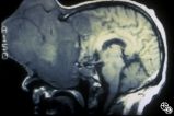
Neuro-Ophthalmic Imaging-MRI | Rosa A. Tang, MD | Aneurisms may result in neuro-ophthalmologic sign and symptoms by direct compression of the afferent or efferent systems or by the secondary effects of hemorrhage. Basilar aneurisms may result in ocular motor deficits such as a unilateral or bilateral third nerve palsy. |
| 152 |
 |
Neuro-Ophthalmic Imaging-MRI | Don Bienfang, MD | A 59-year-old untreated hypertensive man had a sudden onset of vomiting, gait ataxia, dysarthria, and left-sided weakness. The eyes were deviated downward and to the left. (Note position of gaze in 94_11). Extraocular motility was full. There was rotary nystagmus with the fast phase to the left; the... |
| 153 |
 |
Neuro-Ophthalmic Imaging-MRI | Scott Forman, MD | This 23-year-old right-handed man had a history of idiopathic recurrent optic neuritis. The patient presented with acuity of 20/400 OD and 20/100 OS, with a central scotoma OD and a complete temporal defect OS. MRI with fat suppression and gadolinium revealed enhancement of the intracranial nerve an... |
| 154 |
 |
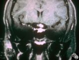
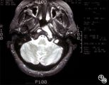
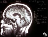
Neuro-Ophthalmic Imaging-MRI | Mitchell J. Wolin, MD | Axial view of Arnold-Chiari malformation on a patient with downbeat nystagmus. Note the presence of the cerebellar tonsils posterior to the caudal medulla. In addition to downbeat nystagmus, Arnold-Chiari malformations can sometimes lead to increased intracranial pressure and papilledema. |
| 155 |
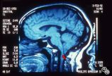 |
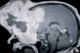
Neuro-Ophthalmic Imaging-MRI | Mitchell J. Wolin, MD | Sagittal view of Arnold-Chiari malformation on a patient with downbeat nystagmus. The compression of the cervicomedullary junction is clearly depicted in the sagittal view. |
| 156 |
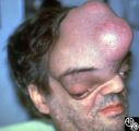 |
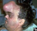
Neuro-Ophthalmic Manifestations of Brain Tumors | Steven Galetta, MD | The patient is a 45-year-old recluse found to harbor this frontal lobe mass. Remarkably, this patient had only mild bilateral optic neuropathies with visual acuities in the 20/25 range. This right disc was mildly swollen and the left mildly pale. He could not fit into the fundus camera for disc phot... |
| 157 |
 |
Neuro-Ophthalmic Manifestations of Brain Tumors | Steven Galetta, MD | The patient is a 45-year-old recluse found to harbor this frontal lobe mass. Remarkably, this patient had only mild bilateral optic neuropathies with visual acuities in the 20/25 range. This right disc was mildly swollen and the left mildly pale. He could not fit into the fundus camera for disc phot... |
| 158 |
 |
Neuro-Ophthalmic Manifestations of Brain Tumors | Jacqueline A. Leavitt, MD | Chordomas of the clivus may result in diplopia due to a sixth nerve palsy. The sixth nerve runs up the clivus and may be the presenting manifestation of the lesion. Pair with Images 97_33, 97_34, and 97_35. |
| 159 |
 |
Neuro-Ophthalmic Manifestations of Brain Tumors | Steven Galetta, MD | The patient is a 45-year-old recluse found to harbor this frontal lobe mass. Remarkably, this patient had only mild bilateral optic neuropathies with visual acuities in the 20/25 range. This right disc was mildly swollen and the left mildly pale. He could not fit into the fundus camera for disc phot... |
| 160 |
 |
Neuro-Ophthalmic Manifestations of Brain Tumors | Steven Galetta, MD | The patient is a 45-year-old recluse found to harbor this frontal lobe mass. Remarkably, this patient had only mild bilateral optic neuropathies with visual acuities in the 20/25 range. This right disc was mildly swollen and the left mildly pale. He could not fit into the fundus camera for disc phot... |
| 161 |
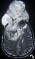 |
Neuro-Ophthalmic Manifestations of Brain Tumors | Steven Galetta, MD | The patient is a 45-year-old recluse found to harbor this frontal lobe mass. Remarkably, this patient had only mild bilateral optic neuropathies with visual acuities in the 20/25 range. This right disc was mildly swollen and the left mildly pale. He could not fit into the fundus camera for disc phot... |
| 162 |
 |
Neuro-Ophthalmic Manifestations of Brain Tumors | Steven Galetta, MD | The patient is a 45-year-old recluse found to harbor this frontal lobe mass. Remarkably, this patient had only mild bilateral optic neuropathies with visual acuities in the 20/25 range. This right disc was mildly swollen and the left mildly pale. He could not fit into the fundus camera for disc phot... |
| 163 |
 |
Neuro-Ophthalmic Manifestations of Brain Tumors | Jacqueline A. Leavitt, MD | Chordomas of the clivus may result in diplopia due to a sixth nerve palsy. The sixth nerve runs up the clivus and may be the presenting manifestation of the lesion. Pair with Images 97_34, 97_35, and 97_36. |
| 164 |
 |
Neuro-Ophthalmic Manifestations of Brain Tumors | Jacqueline A. Leavitt, MD | Chordomas of the clivus may result in diplopia due to a sixth nerve palsy. The sixth nerve runs up the clivus and may be the presenting manifestation of the lesion. Pair with Images 97_33, 97_34, and 97_36. |
| 165 |
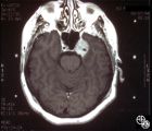 |
Neuro-Ophthalmic Manifestations of Brain Tumors | Jacqueline A. Leavitt, MD | Chordomas of the clivus may result in diplopia due to a sixth nerve palsy. The sixth nerve runs up the clivus and may be the presenting manifestation of the lesion. Imaging: MRI, T-1 axial with contrast. Pair with Images 97_33, 97_34, and 97_36. Anatomy: Clivus, Sixth nerve. Pathology: Sixth nerve p... |
| 166 |
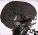 |
Neuro-Ophthalmic Vascular Disease | Mitchell J. Wolin, MD | Carotid cavernous fistulas (CCFs) are connections between the arterial blood flow from the carotid artery system and the cavernous sinus. CCFs may be direct high-flow fistulas or indirect low-flow fistulas. Most direct CCFs are due to trauma. Enlargement of the superior ophthalmic vein may be demons... |
| 167 |
 |
Neuro-Ophthalmic Vascular Disease | Mitchell J. Wolin, MD | Carotid cavernous fistulas (CCFs) are connections between the arterial blood flow from the carotid artery system and the cavernous sinus. CCFs may be direct high-flow fistulas or indirect low-flow fistulas. Most direct CCFs are due to trauma. Enlargement of the superior ophthalmic vein may be demons... |
| 168 |
 |
Neuro-Ophthalmic Vascular Disease | Steven A. Newman, MD | Occlusion of a branch or central retinal artery may result in acute visual loss. The ophthalmoscopic findings are retinal whitening due to ischemic retina in the distribution of the occluded artery. Sparing or selective involvement of cilioretinal artery branches may occur. Patients with a central r... |
| 169 |
 |
Neuro-Ophthalmic Vascular Disease | Steven A. Newman, MD | Occlusion of a branch or central retinal artery may result in acute visual loss. The ophthalmoscopic findings are retinal whitening due to ischemic retina in the distribution of the occluded artery. Sparing or selective involvement of cilioretinal artery branches may occur. Patients with a central r... |
| 170 |
 |
Neuro-Ophthalmic Vascular Disease | Steven A. Newman, MD | Occlusion of a branch or central retinal artery may result in acute visual loss. The ophthalmoscopic findings are retinal whitening due to ischemic retina in the distribution of the occluded artery. Sparing or selective involvement of cilioretinal artery branches may occur. Patients with a central r... |
| 171 |
 |
Neuro-Ophthalmic Vascular Disease | Larry P. Frohman, MD | This 27-year-old woman had no past ocular history and presented with 3 weeks of redness OS that has been treated by the referring doctor as allergic conjunctivitis. She was referred for evaluation when she developed binocular diplopia. Her past medical history included phlebitis and one miscarriage ... |
| 172 |
 |
Neuro-Ophthalmic Vascular Disease | Larry P. Frohman, MD | This 27-year-old woman had no past ocular history and presented with 3 weeks of redness OS that has been treated by the referring doctor as allergic conjunctivitis. She was referred for evaluation when she developed binocular diplopia. Her past medical history included phlebitis and one miscarriage ... |
| 173 |
 |
Neuro-Ophthalmic Vascular Disease | Steven A. Newman, MD | Occlusion of a branch or central retinal artery may result in acute visual loss. The ophthalmoscopic findings are retinal whitening due to ischemic retina in the distribution of the occluded artery. Sparing or selective involvement of cilioretinal artery branches may occur. Patients with a central r... |
| 174 |
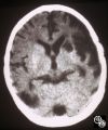 |
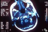
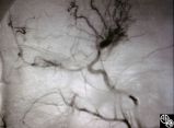
Neuro-Ophthalmic Vascular Disease | Mark J. Kupersmith, MD | A 9-year-old boy had recurrent ischemic episodes that had begun 2 years prior to evaluation. A significant right hemiparesis and a significant speech, learning, and memory disorder were present. His noncontrast axial view CT scan demonstrated multiple cerebral infarcts. Cerebral angiography revealed... |
| 175 |
 |
Neuro-Ophthalmic Vascular Disease | Mark J. Kupersmith, MD | A 9-year-old boy had recurrent ischemic episodes that had begun 2 years prior to evaluation. A significant right hemiparesis and a significant speech, learning, and memory disorder were present. His noncontrast axial view CT scan demonstrated multiple cerebral infarcts. Cerebral angiography revealed... |