A collection of videos relating to the diagnosis and treatment of eye movement disorders. This collection includes many demonstrations of examination techniques.
Dan Gold, D.O., Associate Professor of Neurology, Ophthalmology, Neurosurgery, Otolaryngology - Head & Neck Surgery, Emergency Medicine, and Medicine, The Johns Hopkins School of Medicine.
A collection of videos relating to the diagnosis and treatment of eye movement disorders.
NOVEL: https://novel.utah.edu/
TO
| Title | Description | Type | ||
|---|---|---|---|---|
| 151 |
 |
Bruns Nystagmus (During Video-Oculography) Due to Vestibular Schwannoma | A 25-year-old man with a history of right-sided hearing loss, headaches and imbalance was found to have a right vestibular schwannoma on MRI, and underwent a partial resection and radiotherapy. He denied symptoms of head movement dependent oscillopsia (i.e., suggestive of significant unilateral or b... | Image/MovingImage |
| 152 |
 |
Parinaud's Syndrome in a Man with GBM of the Pineal Gland | 𝗢𝗿𝗶𝗴𝗶𝗻𝗮𝗹 𝗗𝗲𝘀𝗰𝗿𝗶𝗽𝘁𝗶𝗼𝗻: This is a 60-yo-man who presented with diplopia, headaches, and difficulty looking up, and was found to have a mass involving the pineal gland. Biopsy was diagnostic of a GBM. Major features of Parinaud's (dorsal midbrain... | Image/MovingImage |
| 153 |
 |
Congenital Nystagmus | 𝗢𝗿𝗶𝗴𝗶𝗻𝗮𝗹 𝗗𝗲𝘀𝗰𝗿𝗶𝗽𝘁𝗶𝗼𝗻: Presented here are two patients with congenital nystagmus demonstrating characteristic features including: mixed pendular and jerk nystagmus (usually gaze-evoked) waveforms, stays horizontal even in vertical gaze, suppres... | Image/MovingImage |
| 154 |
 |
Test Your Knowledge - Monocular Oscillopsia | 𝗢𝗿𝗶𝗴𝗶𝗻𝗮𝗹 𝗗𝗲𝘀𝗰𝗿𝗶𝗽𝘁𝗶𝗼𝗻: Which of the following associated signs is most likely to be seen in this patient presenting with oscillopsia? A. Optic nerve pallor B. Palatal tremor C. Severe unilateral cataract D. Head bobbing E. Neurovascular contact... | Image/MovingImage |
| 155 |
|
Spontaneous Torsional Nystagmus and Ocular Tilt Reaction | 𝗢𝗿𝗶𝗴𝗶𝗻𝗮𝗹 𝗗𝗲𝘀𝗰𝗿𝗶𝗽𝘁𝗶𝗼𝗻: This is a 70-year-old man who experienced "a delay in focusing" with "some twisting movement" that began about 18 months prior to this video with mild progression over days or weeks. For the same period of time, he experi... | Image/MovingImage |
| 156 |
 |
Ocular Motor Signs in Early Progressive Supranuclear Palsy | 𝗢𝗿𝗶𝗴𝗶𝗻𝗮𝗹 𝗗𝗲𝘀𝗰𝗿𝗶𝗽𝘁𝗶𝗼𝗻: This is a 64-year old man who experienced imbalance and falls (usually backwards) for the last 6 months. He experienced difficulty navigating stairs and had become a messy eater (thought to be in large part due to his ver... | Image/MovingImage |
| 157 |
 |
Trigeminal Neuropathy with Loss of the Corneal Reflex | This is a woman who underwent radiofrequency ablation for left trigeminal neuralgia. Examination demonstrated loss of facial sensation on the left in addition to an absent corneal reflex on the left, consistent with involvement of the V1 (ophthalmic) branch of the trigeminal nerve. When the cornea i... | Image/MovingImage |
| 158 |
 |
Typical Lid Signs (Cogan's Lid Twitch, Lid Hopping, Enhanced Ptosis) in Myasthenia Gravis | 𝗢𝗿𝗶𝗴𝗶𝗻𝗮𝗹 𝗗𝗲𝘀𝗰𝗿𝗶𝗽𝘁𝗶𝗼𝗻: This is a 60-yo-woman with MG who displays typical eyelid signs including Cogan's lid twitch, lid hopping (appreciated during horizontal smooth pursuit in this patient), and enhanced ptosis in accordance with Hering's law... | Image/MovingImage |
| 159 |
 |
Prolonged Lid Twitch in Myasthenia Gravis | This 50-yo-woman with ocular MG demonstrated a spontaneous and particularly prolonged eyelid twitch. | Image/MovingImage |
| 160 |
 |
Internuclear Ophthalmoplegia (INO) in Multiple Sclerosis | 𝗢𝗿𝗶𝗴𝗶𝗻𝗮𝗹 𝗗𝗲𝘀𝗰𝗿𝗶𝗽𝘁𝗶𝗼𝗻: This video includes 3 patients each with a known history of MS found to have unilateral or bilateral INOs on their exam. In the first 2 patients, the INOs are relatively subtle with normal adduction. However, with rapid h... | Image/MovingImage |
| 161 |
 |
Bilateral INOs and Partial 3rd Nerve Palsies | This is a 45-year-old man with progressive ptosis and ophthalmoparesis. 10 years prior to presentation, he experienced diplopia and had a hyperintense lesion involving the medial longitudinal fasciculus (MLF) per report. Over time, he developed bilateral adduction paresis, ptosis and upgaze paresis ... | Image/MovingImage |
| 162 |
 |
Wall-eyed Bilateral INO in Caudal Midbrain Lesion | This is a 30-yo-woman with the relatively acute onset of diplopia. There was a large angle exotropia, very subtle lag of the adducting saccades OD>OS, suggestive of bilateral INOs. This was best seen with rapid horizontal saccades, and a lesion involving bilateral MLFs in the caudal midbrain was dem... | Image/MovingImage |
| 163 |
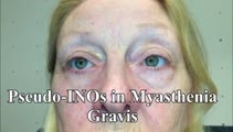 |
Pseudo-INOs in Myasthenia Gravis | This is a 55-yo-woman with an intermittent exotropia who had normal adduction OU, but clear lag of adducting saccades OD>OS with rapid horizontal saccades. This was much more apparent after repeat testing (ie, it was fatigable), and she wound up having ocular MG. | Image/MovingImage |
| 164 |
 |
One-and-a-Half Syndrome, Facial Palsy, and Nystagmus Due to Dorsal Pontine Demyelination | This is a 16-yo-girl with oscillopsia and double vision. Exam showed inability to look to the left with either eye due to left nuclear 6th. There was also a left INO (horizontal gaze palsy + INO = one-and-a-half syndrome) from left MLF involvement and left lower motor neuron facial palsy due to fasc... | Image/MovingImage |
| 165 |
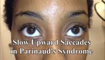 |
Two Patients with Parinaud's Syndrome with Slow Upward Saccades and Normal Upward Range of Movements | Presented here are two patients with Parinaud's syndrome: Patient 1) suffered a hemorrhage of the dorsal midbrain causing slow upward saccades (with convergence retraction nystagmus, but normal vertical range of eye movements), and light-near dissociation, and Patient 2) had a germinoma of the dorsa... | Image/MovingImage |
| 166 |
 |
Parinaud's Syndrome with Impaired Upward Saccades and Otherwise Normal Vertical Eye Movements | This is a 50-yo-man who suffered a dorsal midbrain stroke. Exam demonstrated normal vertical range of eye movements, normal vertical VOR and smooth pursuit, but inability to perform upward saccades. Another feature of Parinaud's syndrome seen on his exam was light-near dissociation (not shown in thi... | Image/MovingImage |
| 167 |
|
Central Positional Vertigo and Nystagmus in a Posterior Fossa Tumor | 𝗢𝗿𝗶𝗴𝗶𝗻𝗮𝗹 𝗗𝗲𝘀𝗰𝗿𝗶𝗽𝘁𝗶𝗼𝗻: This is a 30-year old woman who presented with positional vertigo and vomiting following a concussion related to a car accident 3 months prior. She was initially diagnosed with posterior canal (PC) benign paroxysmal posit... | Image/MovingImage |
| 168 |
 |
Downbeat Nystagmus with Active Horizontal Head Shaking | This is a 70-year-old man who presented with one single complaint for 10 years - if he moved his head too quickly (even one single horizontal head movement to the right or the left), he would experience the abrupt loss of balance and dizziness. His typical episodes were reproducible, and interesting... | Image/MovingImage |
| 169 |
|
Periodic Alternating Nystagmus and Central Head-Shaking Nystagmus from Nodulus Injury | 𝗢𝗿𝗶𝗴𝗶𝗻𝗮𝗹 𝗗𝗲𝘀𝗰𝗿𝗶𝗽𝘁𝗶𝗼𝗻: This is a 35-year-old man who suffered a gunshot wound to his cerebellum. When he regained consciousness days later, he experienced oscillopsia due to periodic alternating nystagmus (PAN). He was started on baclofen 10 mg... | Image/MovingImage |
| 170 |
|
Ocular Motor Signs of Cerebellar Ataxia - Gaze-Evoked Nystagmus, Saccadic Pursuit, and VOR Supression | 𝗢𝗿𝗶𝗴𝗶𝗻𝗮𝗹 𝗗𝗲𝘀𝗰𝗿𝗶𝗽𝘁𝗶𝗼𝗻: This is a 30-year-old woman with a several year long history of imbalance due to cerebellar ataxia of unclear etiology. Seen in this video are common ocular motor signs in patients with advanced cerebellar dysfunction inc... | Image/MovingImage |
| 171 |
 |
Slow Horizontal, Vertical and Oblique Saccades in Spinocerebellar Ataxia Type I | This is a patient presenting with horizontal diplopia who was found to have divergence insufficiency, an esotropia greater at distance than near in the absence of abduction paresis. She also had very slow saccades, more so vertically than horizontally. This is particularly noticeable when asking h... | Image/MovingImage |
| 172 |
 |
Sequelae of Cerebellar Hemorrhage - Gaze-evoked Nystagmus, Alternating Skew Deviation and Palatal Tremor | This is a 75-yo-woman presenting with a gait disorder. Two years prior, she suffered a cerebellar hemorrhage. On examination, there were typical cerebellar ocular motor signs including gaze-evoked nystagmus, choppy smooth pursuit and VOR suppression, and saccadic dysmetria. There was also an alterna... | Image/MovingImage |
| 173 |
 |
Torsional Nystagmus Due to Medullary Pilocytic Astrocytoma | 𝗢𝗿𝗶𝗴𝗶𝗻𝗮𝗹 𝗗𝗲𝘀𝗰𝗿𝗶𝗽𝘁𝗶𝗼𝗻: This is a 30-year-old woman who experienced headaches which led to an MRI and the diagnosis of a right medullary pilocytic astrocytoma, confirmed pathologically. Examination was performed a year after the initial diagnosi... | Image/MovingImage |
| 174 |
 |
Central (Nuclear) 3rd Nerve Palsies | Shown here are two patients with left sided midbrain pathology (hemorrhage and ischemia) which caused damage to the 3rd nucleus. Both of the patients have ipsilateral mydriasis, adduction, supra- and infraduction paresis. Ipsilateral>contralateral ptosis is also present, and localizes to the central... | Image/MovingImage |
| 175 |
 |
Torsional Jerk Nystagmus | Presented here are 3 patients with torsional jerk nystagmus. The first patient presented with vertigo and experienced oscillopsia due to her torsional nystagmus. Pure or predominantly torsional nystagmus is highly suggestive of a central process. Her nystagmus was unidirectional and followed Alexand... | Image/MovingImage |