AAO-NANOS Neuro-Ophthalmology Clinical Collection: Derived from the AAO-NANOS Clinical Neuro-Ophthalmology collection produced on CD. The images are of selected cases from the NANOS teaching slide exchange, and the CD was produced under the direction of Larry Frohman, MD and Andrew Lee, MD.
The American Academy of Ophthalmology (AAO); The North American Neuro-Ophthalmology Association (NANOS).
NOVEL: https://novel.utah.edu/
TO
Filters: Collection: ehsl_novel_aao_nanos
| Title | Description | Subject | ||
|---|---|---|---|---|
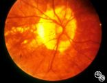
| 126 |
 |
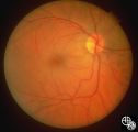
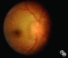
Isolated Congenital Optic Disc Anomalies | An optic pit is a small defect in the optic disc that may be asymptomatic in isolation. Patients may develop an associated serous detachment of the macula. The condition is usually unilateral but may be bilateral. A fluorescein angiogram may demonstrate the serous detachment, and laser photocoagulat... | Optic Pit With Serous Macular Detachment |
| 127 |
 |
Isolated Congenital Optic Disc Anomalies | An optic pit is a small defect in the optic disc that may be asymptomatic in isolation. The pit can be small or large, and central or peripheral. Disease/Diagnosis: Optic Pit. | Optic Pit; Macular Detachment |
| 128 |
 |
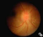
Isolated Congenital Optic Disc Anomalies | Optociliary shunt vessels are venous collaterals that form in response to chronic venous obstruction, shunting the venous blood from the retinal circulation into the choroidal circulation. Although they may be congenital, they may occur in patients with chronic disc edema, following central retinal ... | Optociliary Shunt Vessels |
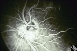
| 129 |
 |
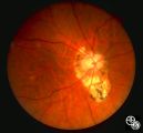
Isolated Congenital Optic Disc Anomalies | Patients with hypoplasia of the optic nerve may have normal or subnormal visual acuity or visual field. The condition may be unilateral or bilateral. Optic nerve hypoplasia is usually idiopathic, but maternal diabetes, or maternal use of anti-epileptic drugs or alcohol are predisposing factors. Opti... | Optic Nerve Hypoplasia; Septo-Optic Dysplasia |
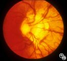
| 130 |
 |
Retinal Coloboma Underneath a Relatively Normal Optic Nerve | Optic nerve colobomas appear as enlarged, white optic discs that are deeply excavated, often with some sapring of the superior rim. They result from an abnormal fusion of the proximal embryonic fissure. Optic nerve colobomas occur unilaterally or bilaterally with a similar frequency and can result i... | Optic Nerve Coloboma |
| 131 |
 |
Isolated Congenital Optic Disc Anomalies | Patients with midline closure defects may exhibit abnormalities in the optic nerve, choroid, retinal pigment epithelium or retina. Anterior closure defects may result in colobomas of the structures of the anterior segment, such as the iris. Disease/Diagnosis: Coloboma. | Optic Nerve Coloboma |
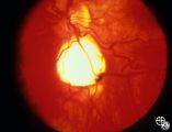
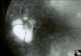
| 132 |
 |
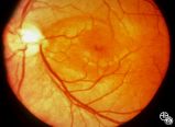
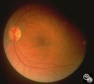
Peripapillary Staphyloma | Patients with ectasia of the outer layers of the eye may exhibit a posterior protrusion that appears on funduscopy as an area of deep excavation of the retina (posterior staphyloma). When it occurs around the optic disc, as in this case, it is termed a peripapillary staphyloma. This may occur in ass... | Staphyloma |
| 133 |
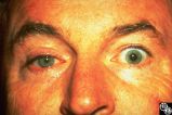 |
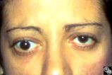
Migraine Syndrome | The image shows a patient with cluster headache and eye displaying Horner's syndrome. | Cluster Headache |
| 134 |
 |
Neuro-Ophthalmic Imaging-MRI | Axial view of Arnold-Chiari malformation on a patient with downbeat nystagmus. Note the presence of the cerebellar tonsils posterior to the caudal medulla. In addition to downbeat nystagmus, Arnold-Chiari malformations can sometimes lead to increased intracranial pressure and papilledema. | Arnold-Chiari Malformation; Chiari Malformation; Inferior Tonsillar Herniation; Downbeat Nystagmus |
| 135 |
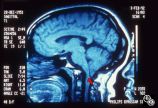 |
Neuro-Ophthalmic Imaging-MRI | Sagittal view of Arnold-Chiari malformation on a patient with downbeat nystagmus. The compression of the cervicomedullary junction is clearly depicted in the sagittal view. | Arnold-Chiari Malformation; Chiari Malformation; Inferior Tonsillar Herniation; Downbeat Nystagmus |
| 136 |
 |
Neuro-Ophthalmic Vascular Disease | This 32-year-old woman was referred with a history of 4 days of loss of vision OD. She had a history of manic depressive illness and IV drug abuse; she had been HIV tested 4 weeks before and was negative. She said she last injected cocaine 5 days before being seen, the night before she awoke with th... | Saturday Night Retinopathy |
| 137 |
 |
Neuro-Ophthalmic Vascular Disease | This 32-year-old woman was referred with a history of 4 days of loss of vision OD. She had a history of manic depressive illness and IV drug abuse; she had been HIV tested 4 weeks before and was negative. She said she last injected cocaine 5 days before being seen, the night before she awoke with th... | Saturday Night Retinopathy |
| 138 |
 |
Neuro-Ophthalmic Vascular Disease | This 32-year-old woman was referred with a history of 4 days of loss of vision OD. She had a history of manic depressive illness and IV drug abuse; she had been HIV tested 4 weeks before and was negative. She said she last injected cocaine 5 days before being seen, the night before she awoke with th... | Saturday Night Retinopathy |
| 139 |
 |
Neuro-Ophthalmic Vascular Disease | This 32-year-old woman was referred with a history of 4 days of loss of vision OD. She had a history of manic depressive illness and IV drug abuse; she had been HIV tested 4 weeks before and was negative. She said she last injected cocaine 5 days before being seen, the night before she awoke with th... | Saturday Night Retinopathy |
| 140 |
 |
Neuro-Ophthalmic Vascular Disease | This 32-year-old woman was referred with a history of 4 days of loss of vision OD. She had a history of manic depressive illness and IV drug abuse; she had been HIV tested 4 weeks before and was negative. She said she last injected cocaine 5 days before being seen, the night before she awoke with th... | Saturday Night Retinopathy |
| 141 |
 |
Systemic Disorders With Optic Nerve and Retinal Findings | At age 41, in 1984, this woman, who grew up in the Ohio River Valley, had 3 days of ocular pain OD, and her vision declined to 20/80 OD she has had no visual changes since, nor has she had any other neurologic symptoms. The ""presumed"" diagnosis is optic neuropathy in presumed ocular histoplasmosis... | Presumed Ocular Histoplasmosis |
| 142 |
 |
Systemic Disorders With Optic Nerve and Retinal Findings | This is a 32-year-old HIV-positive man with anterior uveitis, vitritis, and bilateral papillitis from syphilis. With intravenous penicillin treatment, the optic discs and vision returned to normal. | Syphilis |
| 143 |
 |
Systemic Disorders With Optic Nerve and Retinal Findings | This is a 32-year-old HIV-positive man with anterior uveitis, vitritis, and bilateral papillitis from syphilis. With intravenous penicillin treatment, the optic discs and vision returned to normal. | Syphilis |
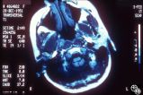
| 144 |
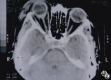 |
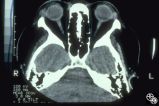
Neuro-Ophthalmic Imaging-CT Scan | This 70-year-old woman sustained traumatic optic neuropathy in a motor vehicle accident. Note the funnel-shaped hemorrhage within the optic nerve sheath just posterior to the globe. | Traumatic Optic Neuropathy |
| 145 |
 |
Neuro-Ophthalmic Imaging-CT Scan | This 39-year-old woman's initial sign was painless, progressive, symmetric ptosis OU, without diurnal variation, that manifested when she was age 17 living in the Dominican Republic. At that time, she had no diplopia or systemic signs. She had no family history of ocular or muscle disease, and no ot... | Traumatic Optic Neuropathy |
| 146 |
 |
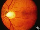
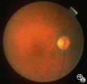
Isolated Congenital Optic Disc Anomalies | This 63-year-old man with amblyopia OD was seen for a question of ischemic optic neuropathy with a pale, swollen disc OD. The correct diagnosis is an exophytic capillary angioma of the optic nerve head. Disease/Diagnosis: Capillary Angioma. | Capillary Angioma |
| 147 |
 |
Isolated Congenital Optic Disc Anomalies | This 63-year-old man with amblyopia OD was seen for a question of ischemic optic neuropathy with a pale, swollen disc OD. The correct diagnosis is an exophytic capillary angioma of the optic nerve head. Disease/Diagnosis: Capillary Angioma. | Capillary Angioma |
| 148 |
 |
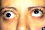
Optic Tract Syndrome Due to Carotid Artery Dolichoectasia | This 43-year-old man was referred for evaluation of 6 months of visual loss OU. In retrospect, he had noticed increasing difficulty with his tennis game dating back over 3 years, as balls would pass him unexpectedly when hit to his backhand (left) side. The patient did not think this was progressive... | Dolichoectasia |
| 149 |
 |
Optic Tract Syndrome Due to Carotid Artery Dolichoectasia | This 43-year-old man was referred for evaluation of 6 months of visual loss OU. In retrospect, he had noticed increasing difficulty with his tennis game dating back over 3 years, as balls would pass him unexpectedly when hit to his backhand (left) side. The patient did not think this was progressive... | Dolichoectasia |
| 150 |
 |
Optic Tract Syndrome Due to Carotid Artery Dolichoectasia | This 43-year-old man was referred for evaluation of 6 months of visual loss OU. In retrospect, he had noticed increasing difficulty with his tennis game dating back over 3 years, as balls would pass him unexpectedly when hit to his backhand (left) side. The patient did not think this was progressive... | Dolichoectasia |