Collection of materials relating to neuro-ophthalmology as part of the Neuro-Ophthalmology Virtual Education Library.
NOVEL: https://novel.utah.edu/
TO
- NOVEL224
| Title | Creator | Description | Subject | ||
|---|---|---|---|---|---|
| 126 |
 |
Grant Pieces - Research Grants | Silvia Sörensen, PhD | Lecture describing the parts of a research grant, including using human subjects. | Grant Writing |
| 127 |
 |
My DEI Journey: Lessons Learned | Ore-ofe Adesina, MD | In this lecture, Dr. Adesina reviews his DEI journey in ophthalmology and the lessons of diversity, equity and inclusion he has learned along the way. He discusses why diversity, equity, and inclusion are all needed for a state of inclusive well-being, why representation matters for patient care and... | Ophthalmology; Diversity, Equity, Inclusion; Representation; Social Determinants of Health |
| 128 |
 |
Professionalism and Communication Skills | Karl C. Golnik, MD, MEd | Lecture covering professionalism and communication skills. | Professionalism; Communication Skills |
| 129 |
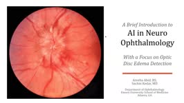 |
A Brief Introduction to AI in Neuro-ophthalmology | Areeba Abid, BS; Sachin Kedar, MD | In this video, we describe the basics of artificial intelligence and machine learning as applicable to clinical neuro-ophthalmologists. We use the example from a recent publication, where AI software was used to detect optic disc edema in fundus images. | Artificial Intelligence; Machine Learning |
| 130 |
 |
Bony Anatomy of the Orbit | Aakash Patel; Amanda Henderson, MD | Narrated presentation on the bony anatomy of the orbit. | Bony Anatomy; Orbit |
| 131 |
 |
Physiology of the Uvea | Aakash Patel; Amanda Henderson, MD | Overview of the physiology of the uvea. | Uvea; Physiology |
| 132 |
 |
Ocular Surface, Cornea, & Lens | Sari Yordi, MD | Video lecture on the anatomy of the ocular surface, cornea, and lens. | Ocular Surface; Cornea; Lens |
| 133 |
 |
Lacrimal Pathways: Anatomy and Physiology | Sari Yordi, MD | Video lecture covering anatomy and physiology of the lacrimal pathways. | Lacrimal Pathways |
| 134 |
 |
Aqueous and Vitreous Humor | Sari Yordi, MD | Narrated lecture on the aqueous and vitreous humor. | Aqueous; Vitreous Humor |
| 135 |
 |
Physiology of Intraocular Pressure | Aakash Patel; Amanda Henderson, MD | Overview of the physiology of intraocular pressure. | Intraocular Pressure; Physiology |
| 136 |
 |
Optic Chiasm | Yesha Shah, BSA, BBA; Amanda Henderson, MD | Overview of the anatomy of the optic chiasm. | Optic Chiasm; Anatomy |
| 137 |
 |
Control of the Eye Movements: Neuroanatomy Video Lab - Brain Dissections | Suzanne S. Stensaas, PhD | Disturbances in eye movements can provide important clues for localization of neurological damage. The role of the frontal eye fields in horizontal gaze is stressed. The need to coordinate cranial nerves on both sides of the brain stem introduces the medial longitudinal fasciculus and its role in co... | Medial Longitudinal Fasciculus; Third Cranial Nerve; Sixth Cranial Nerve; Internuclear Ophthalmoplegia; Nystagmus; Brain; Dissection |
| 138 |
 |
Optic Nerve Sheath Fenestration | Raed Behbehani, MD | Optic nerve sheath fenestration is performed to manage papilledema causing progressive loss of vision , due to raised intracranial pressure from Idiopathic Intracranial Hypertension or Cerebral Venous Sinus Thrombosis. The procedure is usually performed in cases of severe visual field loss or when m... | Optic Nerve Sheath Fenestration |
| 139 |
 |
Superonasal Transconjunctival Optic Nerve Sheath Decompression (stONSD) | Kevin E. Lai, MD; Kenneth C. Lao, MD; Peter L. Hildebrand, MD; Bradley K. Farris, MD | This video demonstrates the surgical technique and outcomes of a modified medial transconjunctival approach to optic nerve sheath decompression (ONSD). Disease/Diagnosis: Papilledema; Idiopathic Intracranial Hypertension (IIH). | Superonasal Transconjunctival Optic Nerve Sheath Decompression (ONSD); Surgical Technique |
| 140 |
 |
Lateral Orbitotomy with Bone Window | Richard C. Allen, MD, PhD, FACS | Narrated video of lateral orbitotomy with bone removal for improved orbital decompression. | Orbitotomy; Orbital Decompression |
| 141 |
 |
Lateral Orbitotomy #3 | Richard C. Allen, MD, PhD, FACS | Narrated video of lateral orbitotomy and biopsy of presumed benign cavernous hemangioma. | Orbital Biopsy; Orbitotomy |
| 142 |
 |
Lateral Orbitotomy #2 | Richard C. Allen, MD, PhD, FACS | Narrated video of lateral orbitotomy and biopsy of presumed orbital inflammatory condition. | Orbital Biopsy; Orbitotomy |
| 143 |
 |
Comparison of the Motor Systems: Neuroanatomy Video Lab - Brain Dissections | Suzanne S. Stensaas, PhD | A comparison of the three major motor systems focuses on categorizing motor problems as corticospinal tract, cerebellar, or basal ganglia. It begins with 12 minutes of gross anatomical structures and pathway review followed by clinical video clips demonstrating clinical features of disease of each o... | Corticospinal Tract; Cerebellar; Basal Ganglia; Motor Systems; Brain; Dissection |
| 144 |
 |
Basal Ganglia: Neuroanatomy Video Lab - Brain Dissections | Suzanne S. Stensaas, PhD | Structures involved in involuntary movements are shown on models, in animations, and on gross coronal and axial sections. Diagrams show the cortex-to-cortex loop and the nigrostriatal pathway. The direct and indirect pathways are shown. The direct pathway facilitates movement while the indirect path... | Basal Ganglia; Involuntary Movements; Brain; Dissection |
| 145 |
 |
Cortical Localization: Neuroanatomy Video Lab - Brain Dissections | Suzanne S. Stensaas, PhD | The lobes of the brain are defined together with their major functions. The visual field representation in the occipital lobe is explained with a diagram. Speech areas and the major types of aphasia are discussed in the dominant hemisphere and parietal lesions of neglect and spatial orientation are ... | Cortical Localization; Brain; Dissections |
| 146 |
 |
Introduction: Neuroanatomy Video Lab - Brain Dissections | Suzanne S. Stensaas, PhD | The regions and lobes of the brain are identified along with some of the nerves and vessels. The basic functions of the cortex of each lobe are introduced along with principal sulci and gyri. The importance of the left hemisphere for language and the temporal lobe in memory are mentioned along with ... | Brain; Dissections |
| 147 |
 |
Brain Stem & Reflexes: Neuroanatomy Video Lab - Brain Dissections | Suzanne S. Stensaas, PhD | The cranial nerves are reviewed again on a specimen with vessels. Next, landmarks on gross brain stem sections are shown. Stressed are the three reflexes associated with each of the three levels: pupillary, corneal and gag reflexes and their associated cranial nerves. Finally cross sections of myeli... | Brain Stem; Reflexes |
| 148 |
 |
Cerebral Circulation: Neuroanatomy Video Lab - Brain Dissections | Suzanne S. Stensaas, PhD | The major vessels of the anterior and posterior circulation are demonstrated along with the Circle of Willis on both a model and in an animation. The distribution of the three major cerebral arteries is demonstrated along with the concept of a watershed zone. A gross specimen with good vessels is al... | Cerebral Circulation; Brain; Dissections |
| 149 |
 |
Sixth nerve palsy or not? (Video) | Vivian Paraskevi Douglas; Konstantinos Douglas; Nurhan Torun | Here in we present a case of a 72-yearold Caucasian female with remote history of breast cancer who presented with diplopia in the right gaze over the past several months. There had been no improvement after a 5-day course of steroids by an outside ophthalmologist. On neuro-ophthalmic examination, t... | Abduction deficit; Diplopia; Orbital imaging |
| 150 |
 |
Cerebellum: Neuroanatomy Video Lab - Brain Dissections | Suzanne S. Stensaas, PhD | The gross features of the cerebellum are shown. The three peduncles are demonstrated, noting their input and output to and from the cerebellum. Emphasis is given to symptoms of cerebellar disease appearing on the same side of the body. Special emphasis is given to cortical-cerebellar connections, st... | Cerebellum; Cortical-Cerebellar Connection; Brain; Dissection |