Collection of materials relating to neuro-ophthalmology as part of the Neuro-Ophthalmology Virtual Education Library.
NOVEL: https://novel.utah.edu/
TO
- NOVEL226
| Title | Creator | Description | Subject | ||
|---|---|---|---|---|---|
| 126 |
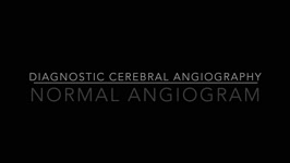 |
Normal Angiogram | Justin Gibson, MD; Charles Prestigiacomo, MD | Example of a normal diagnostic cerebroangiogram. | Angiogram |
| 127 |
 |
Normal Light Reflex and Relative Afferent Pupillary Defect (RAPD) | Marshall Huang, 4th Year Medical Student | A Relative Afferent Pupillary Defect is an examination finding in patients who have an asymmetric pupillary reaction to light when it is shined back and forth between the two eyes. It is most commonly a sign of asymmetric optic nerve disease or damage but can also present in widespread asymmetric r... | Light Reflex; RAPD |
| 128 |
 |
Nystagmus Elicitation Techniques | Jorge C. Kattah, MD | An examination of the patient days or weeks after the acute event requires fixation block, and a variety of techniques, known as nystagmus elicitation maneuvers to detect the recent vestibular imbalance. | Nystagmus |
| 129 |
 |
OJS Author Tutorial | A. Roylance | Tutorial for testing new OJS system for reviews of NOVEL submissions. | OJS Review System |
| 130 |
 |
OJS Reviewer Tutorial | A. Roylance | Tutorial for testing new OJS system for reviews of NOVEL submissions. | OJS Review System |
| 131 |
 |
Ocular Lateropulsion Left AICA Stroke | Jorge C Kattah, MD | 82 year-old patient with basilar artery stenosis, she developed an acute left AICA stroke. On examination within 24 hours from symptom onset, she had primary gaze, unidirectional, right beat nystagmus and a positive left head impulse test. Brief periods of eyelid closure were associated with a h-... | Ocular Lateropulsion; AICA Stroke |
| 132 |
 |
Ocular Neuromyotonia | Raed Behbehani, MD | Ocular Neuromytonia is a characterised by by paroxysmal tonic contraction of the extraocular muscles supplied by the oculomotor nerve. It is has been reported after cranial radiation therapy, especially to the sellar-parasellar region and from compressive lesions such tumours or aneurysms. The patho... | Ocular Neuromyotania |
| 133 |
 |
Ocular Neuromyotonia | Khawla Elnour; Amanda Henderson, MD | An overview of complications of uveitis. | Uveitis; Neuromyotonia |
| 134 |
 |
Ocular Neuromyotonia Video | Bashaer Aldhahwani, MD; Joshua Pasol, MD | A video demonstrates ocular neuromyotonia in the left eye of a patient with a history of cranial radiation of parasellar mass. Ocular neuromyotonia (ONM) is a rare ocular motor disorder characterized by intermittent, tonic spasms of one or more of the extraocular muscles, resulting in strabismus and... | Ocular Neuromyotonia |
| 135 |
 |
Ocular Surface, Cornea, & Lens | Sari Yordi, MD | Video lecture on the anatomy of the ocular surface, cornea, and lens. | Ocular Surface; Cornea; Lens |
| 136 |
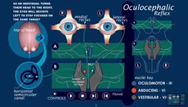 |
Oculocephalic Reflex Animation | Suzanne S. Stensaas, PhD; Quentin Roper | Animation describing the vestibuloocular reflex. | Vestibuloocular Reflex; Vestibular Ocular System |
| 137 |
 |
Oculopalatal Tremor | Raed Behbehani, MD | This is a usually vertical, pendular nystagmus associated with synchronous rhythmic movement of the palate, developing months after a severe brain stem stroke. The stroke involves the dentato-rubro-olivary tract (Mollaret's triangle). MRI can show hypertrophy of the inferior olivary nucleus in the m... | Oculopalatal Tremor |
| 138 |
 |
Olfactory System: Neuroanatomy Video Lab - Brain Dissections | Suzanne S. Stensaas, PhD | Beginning with the location of the sensory cells within the skull the axons are traced into the cranial cavity. Demonstration of the olfactory bulb, olfactory tract and it termination in the forebrain and temporal lobe are indicated. Trauma and meningiomas can produce loss of small (anosmia). Degene... | Olfactory System; Olfactory Bulb; Anosmia; Brain; Dissection |
| 139 |
 |
One and a Half Syndrome Following Resection of a Posterior Fossa Epidermoid Cyst | Christine Xu; Claire Basco, MS, NP-C; Kiarash Shahlaie; Yin Allison Liu | This is a case of One and a Half Syndrome following resection of a posterior fossa epidermoid cyst. A 31-year-old male initially presented with left facial droop and bilateral ptosis, and a "down and out" gaze of the left eye. He underwent imaging and was diagnosed with an epidermoid cyst located in... | Abducens Nucleus; Epidermoid Cyst; Extraocular Movements; Gaze Palsy; Internuclear Ophthalmoplegia; Medial Longitudinal Fasciculus; Neurosurgery; One and a Half Syndrome |
| 140 |
 |
Optic Chiasm | Yesha Shah, BSA, BBA; Amanda Henderson, MD | Overview of the anatomy of the optic chiasm. | Optic Chiasm; Anatomy |
| 141 |
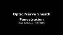 |
Optic Nerve Sheath Fenestration | Raed Behbehani, MD | Optic nerve sheath fenestration is performed to manage papilledema causing progressive loss of vision , due to raised intracranial pressure from Idiopathic Intracranial Hypertension or Cerebral Venous Sinus Thrombosis. The procedure is usually performed in cases of severe visual field loss or when m... | Optic Nerve Sheath Fenestration |
| 142 |
 |
Optic Neuropathy: A Recipe for Blindness | Karim Kozhaya, MD; Alaa Bou Ghannam, MD; Alfredo Sadun, MD, PhD | An epidemic of blindness and peripheral neuropathy struck Cuba in the early 90s. By the end of 1993, 7% of the population was affected. Most patients were men and presented with sub-acute, painless, bilateral loss of vision. The etiology of the disease pondered local and international scientists, es... | Cuban Epidemic Optic Neuropathy; Leber's Hereditary Optic Neuropathy; Mitochondrial Insufficiency; Nutritional Optic Neuropathy; Pale Optic Nerve |
| 143 |
 |
Optic Tract Syndrome Secondary to Lacunar Infarction | Justin J. Grassmeyer, PhD; Kenan Xiao, MD; Jason T. Helvey, MD; Sachin Kedar, MD | Lesions of the optic tract produce a characteristic triad of clinical findings: contralateral homonymous hemianopia, contralateral relative afferent pupillary defect, and bilateral optic disc atrophy. This case describes clinical features, radiological findings, and optical imaging characteristics f... | Optic Tract Syndrome; Optic Tract; Lacunar Infarction; Homonymous Hemianopia |
| 144 |
 |
Optical Coherence Tomography Angiography | David Zhao; Amanda Henderson, MD | Video presentation covering a thorough overview of Optical Coherence Tomography Angiography (OCTA). | Optical Coherence Tomography Angiography; OCTA |
| 145 |
 |
Orientation: The Planes of the Brain: Neuroanatomy Video Lab - Brain Dissections | Suzanne S. Stensaas, PhD | Terms such as anterior, posterior, inferior and superior are introduced with respect to the hemispheres as well as the brain stem. Terms such as rostral and caudal or dorsal and ventral can mean different things in different areas. Sections in three planes (frontal, axial, and sagittal) are demonstr... | Frontal; Axial; Sagittal; Brain; Dissection |
| 146 |
 |
Other Special Situations | John Pula, MD | Introduction to examinations in special situations. | Exams |
| 147 |
 |
Overview of Medical Malpractice, Torts, and Tort Reform | Sarah Jacober; Sean Gratton | This is a brief narrated powerpoint, which serves as an introduction to the basics of medical malpractice. The definition and history of medical malpractice are explored. The relationship between medical malpractice and tort law are explained, and tort reform is introduced. | Lawsuit; Malpractice; Medical Malpractice; Medicolegal; Tort Reform; Torts |
| 148 |
 |
Panoptic Ophthalmoscope | Amrita D. Vuppala, MD | Demonstration of using the panoptic ophthalmoscope in examinations. | Panoptic Ophthalmoscope |
| 149 |
 |
Parinaud Syndrome | Raed Behbehani, MD | Parinaud syndrome, as called dorsal midbrain syndrome, is due to dorsal midbrain lesions from compression (e.g., a tumor), demyelination, or ischemia. The syndrome is characterized by limitation of upward gaze, convergence retraction nystagmus, light near dissociation, and lid retraction (Collier's ... | Dorsal Mibrain Syndrome; Parinaud's Syndrome |
| 150 |
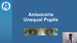 |
Patient Portal: Anisocoria | Nagham Al-Zubidi, MD | Anisocoria is a medical term for unequal pupil size. Normally our pupils are relatively the same size. While small differences in pupil size are normal and can even come and go (physiologic anisocoria), constant and significant differences in pupil sizes may be a sign of damage to the brain or the n... | Anisocoria; Horner Syndrome; 3rd Cranial Nerve Palsy; Adie Tonic Pupil |