Best known for his world-renowned neuro-ophthalmology unit based at the University of California, San Francisco, William Hoyt, MD collected here more than 850 of his best images covering a wide range of disorders.
William F. Hoyt, MD, Professor Emeritus of Ophthalmology, Neurology and Neurosurgery, Department of Ophthalmology, University of California, San Francisco.
NOVEL: https://novel.utah.edu/
TO
Filters: Collection: "ehsl_novel_wfh"
| Title | Description | Type | ||
|---|---|---|---|---|
| 126 |
 |
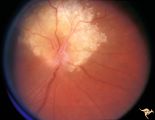
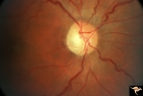
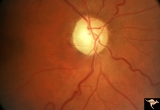
Tuberous Sclerosis | Tuberous Sclerosis. Has two astrocytic hamartomas. One is calcified. Right eye. Pair with R1_D4b. Anatomy: Optic disc; Retina. Pathology: Astrocytic hamartoma. Disease/Diagnosis: Tuberous sclerosis. Clinical: No visual symptoms. | Image |
| 127 |
 |
Von Hippel Lindau Disease (Retinal Hemangioblastoma) | Von Hippel Lindau Disease with large peripheral retinal hemangioblastoma. View of the tumor. Larger artery entering and the vein leaving the tumor are evidence of rapid arteriovenous shunting within the tumor. Group with R1_C3b, R1_C3a, R1_C3d. Anatomy: Retina. Pathology: Hemangioblastoma. Disease/... | Image |
| 128 |
 |
Von Hippel Lindau Disease (Retinal Hemangioblastoma) | Von Hippel Lindau Disease with large retinal hemangioblastoma. Continued view of the arteriole and venous channels leading to the tumor. Group with R1_C3a, R1_C3c, R1_C3d. Anatomy: Retina. Pathology: Hemangioblastoma. Disease/Diagnosis: Von Hippel Lindau disease. Clinical: No visual symptoms. | Image |
| 129 |
 |
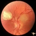
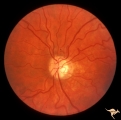
Von Hippel Lindau Disease (Hemangioblastoma of the Optic Disc) | Von Hippel Lindau Disease with a retinal hemangioblastoma on her optic disc. Anatomy: Optic disc. Pathology: Hemangioblastoma. Disease/Diagnosis: Von Hippel Lindau disease. Clinical: No visual symptoms. Patient had cerebellar ataxia. Imaging: R1_C1b is Arteriogram showing hemangioblastoma of the cer... | Image |
| 130 |
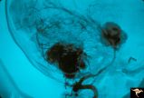 |
Von Hippel Lindau Disease (Hemangioblastoma of the Optic Disc) | Von Hippel Lindau Disease; Arteriogram showing hemangioblastoma of the cerebellum and midbrain. Anatomy: Brain. Pathology: Hemangioblastoma. Disease/Diagnosis: Von Hippel Lindau disease. Clinical: No visual symptoms. Patient had cerebellar ataxia. Imaging: Arteriogram showing hemangioblastoma of the... | Image |
| 131 |
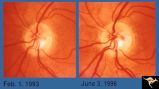 |
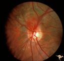
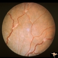
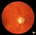
Von Hippel Lindau Disease | Von Hippel Lindau lesion on optic disc showing minimal increase in size over three year interval. Anatomy: Optic disc. Pathology: Hemangioblastoma. Disease/Diagnosis: Von Hippel Lindau disease. Clinical: Patient other eye was removed for hemangioblastoma. He has numerous hemangioblastomas of his spi... | Image |
| 132 |
 |
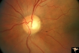
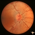
Tuberous Sclerosis | Tuberous Sclerosis. Astrocytic hamartoma of the optic disc. Anatomy: Optic disc. Pathology: Astrocytic hamartoma. Disease/Diagnosis: Tuberous sclerosis. Clinical: No visual symptoms. | Image |
| 133 |
 |
Cerebroretinal Microangiopathy (Susac Syndrome) | Retinal signs of Susac's Syndrome in acute phase consist of areas of retinal artery infarction from branch retinal artery occlusions. This fundus shows two area of retinal infarction from occlusion of both superior and inferior branch retinal arterioles. Anatomy: Retina. Pathology: Microangiopathy... | Image |
| 134 |
 |
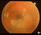
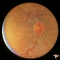
C302 Nodular Papillopathies (Sarcoid) | Perivenous Inflammatory Cuffing in a Patient with Proven Sarcoid. Left eye. Pair with C3_01. Anatomy: Retina. Pathology: Axoplasmic stasis due to sarcoid infiltration with retinal venous exudation? Disease/ Diagnosis: Sarcoid papillopathy with perivenous inflammatory disease. Clinical: Unknown? | Image |
| 135 |
 |
C306 Nodular Papillopathies (Sarcoid) | Lumpy disc swelling with retinal folds and a macular star in a patient with sarcoid. Presentation in October 1983. Same patient as C3_05 and C3_07. Anatomy: Optic disc; Retina. Pathology: Axoplasmic stasis due to sarcoid infiltration. Disease/ Diagnosis: Axoplasmic stasis due to sarcoid infiltration... | Image |
| 136 |
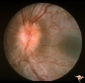 |
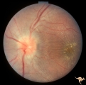
C301 Nodular Papillopathies (Sarcoid) | Disc swelling. Sarcoid papillopathy. Note infiltrative nodule at 9:00 on the disc.The patient had proven sarcoid. Perivenous inflammatory cuffing visible on image C3_02. Right eye. Pair with C3_02. Anatomy: Optic disc; Retina. Pathology: Axoplasmic stasis due to sarcoid infiltration. Disease/ Diagn... | Image |
| 137 |
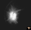 |
D107 Disc Edema with Systemic Lupus | Late stage Flourescein angiogram showing flourescein leakage on the disc and around the neighboring vessels. Note this amount of edema could not be appreciated in the colored fundus image D1_05. Same patient as D1_06 an D1_05. Anatomy: Optic disc. Pathology: Axoplasmic stasis due to vasculitis (Lupu... | Image |
| 138 |
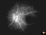 |
D106 Disc Edema with Systemic Lupus | Flourescein angiogram shows evidence of vascular papillopathy. (Lupus) Same patient as D1_05 an D1_07. Anatomy: Optic disc. Pathology: Axoplasmic stasis due to vasculitis (Lupus). Disease/ Diagnosis: Lupus papillopathy. | Image |
| 139 |
 |
IE04 Acute Leber Optic Neuropathy | Microangiopathy without visual loss in a patient with acute Leber's optic neuropathy in the left eye. Pair with IE_05. Anatomy: Optic disc. Pathology: Optic neuropathy. Disease/ Diagnosis: Leber's optic neuropathy. Clinical: Central vision loss. | Image |
| 140 |
 |
IE05 Acute Leber Optic Neuropathy | Patient has just begun to lose vision in his left eye due to Leber's optic neuropathy. Pair with IE_04. Anatomy: Optic disc. Pathology: Optic neuropathy. Disease/ Diagnosis: Leber's optic neuropathy. Clinical: Central vision loss. | Image |
| 141 |
 |
IE11 Subacute Leber Optic Neuropathy | Subacute Leber's Optic Neuropathy with distinct temporal wedge pallor and adjacent microangiopathy. 1973. Anatomy: Optic disc. Pathology: Optic neuropathy. Disease/ Diagnosis: Leber's optic neuropathy. Clinical: Blindness. | Image |
| 142 |
 |
End Stage Leber Optic Neuropathy | End stage Leber's Optic Neuropathy. Severe diffuse pallor. Left eye. Pair with 15a. Anatomy: Optic disc. Pathology: Optic neuropathy. Disease/ Diagnosis: Leber's optic neuropathy. Clinical: Blindness. | Image |
| 143 |
 |
IE12a Acute Leber Optic Neuropathy | Subacute stage of Leber's Optic Neuropathy showing microangiopathy showing temporal pallor. Atrophy more advanced in left eye (12b) 1971, right eye. Anatomy: Optic disc. Pathology: Optic neuropathy. Disease/ Diagnosis: Leber's optic neuropathy. Clinical: Blindness. | Image |
| 144 |
 |
IE12b Subacute Leber Optic Neuropathy | Subacute stage of Leber's Optic Neuropathy showing microangiopathy showing temporal pallor. Atrophy more advanced in left eye. 1971, left eye. Anatomy: Optic disc. Pathology: Optic neuropathy. Disease/ Diagnosis: Leber's optic neuropathy. Clinical: Blindness. | Image |
| 145 |
 |
IE13a End Stage Leber Optic Neuropathy | End stage Leber's Optic Neuropathy. Note modest arteriolar narrowing. Note also the generalized pallor of the disc. Microangiopathy is no longer visible. Right eye. Pair with 13b. Anatomy: Optic disc. Pathology: Optic neuropathy. Disease/ Diagnosis: Leber's optic neuropathy. Clinical: Blindness. | Image |
| 146 |
 |
IE13b End Stage Leber Optic Neuropathy | End stage Leber's Optic Neuropathy. Note modest arteriolar narrowing. Note also the generalized pallor of the disc. Microangiopathy is no longer visible. Left eye. Pair with 13a. Anatomy: Optic disc. Pathology: Optic neuropathy. Disease/ Diagnosis: Leber's optic neuropathy. Clinical: Blindness. | Image |
| 147 |
 |
IE14a End Stage Leber Optic Neuropathy | End stage Leber's Optic Neuropathy. Dense temporal pallor. Microangiopathy is absent. Right eye. Pair with 14b. Anatomy: Optic disc. Pathology: Optic neuropathy. Disease/ Diagnosis: Leber's optic neuropathy. Clinical: Blindness. | Image |
| 148 |
 |
IE14b End Stage Leber Optic Neuropathy | End stage Leber's Optic Neuropathy. Dense temporal pallor. Microangiopathy is absent. Left eye. Pair with 14a. Anatomy: Optic disc. Pathology: Optic neuropathy. Disease/ Diagnosis: Leber's optic neuropathy. Clinical: Blindness. | Image |
| 149 |
 |
IE15a End Stage Leber Optic Neuropathy | End stage Leber's Optic Neuropathy. Severe diffuse pallor. Right eye. Pair with 15b. Anatomy: Optic disc. Pathology: Optic neuropathy. Disease/ Diagnosis: Leber's optic neuropathy. Clinical: Blindness. | Image |
| 150 |
 |
IE03 Acute Leber Optic Neuropathy | Acute stage of Leber optic neuropathy with microangiopathy and peripapillary nerve fiber layer thickening. The temporal nerve fiber layer is already showing atrophy. Central vision is grossly reduced. 1971. Anatomy: Optic disc. Pathology: Optic neuropathy. Disease/ Diagnosis: Leber's optic neuropath... | Image |