AAO-NANOS Neuro-Ophthalmology Clinical Collection: Derived from the AAO-NANOS Clinical Neuro-Ophthalmology collection produced on CD. The images are of selected cases from the NANOS teaching slide exchange, and the CD was produced under the direction of Larry Frohman, MD and Andrew Lee, MD.
The American Academy of Ophthalmology (AAO); The North American Neuro-Ophthalmology Association (NANOS).
NOVEL: https://novel.utah.edu/
TO
Filters: Collection: ehsl_novel_aao_nanos
| Title | Description | Subject | ||
|---|---|---|---|---|
| 101 |
 |
Motility Disturbances | This patient displays a posttraumatic left fourth nerve palsy sustained after having struck her head on the dashboard. | Trochlear Palsy; Fourth (Trochlear) |
| 102 |
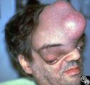 |
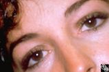
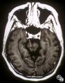
Neuro-Ophthalmic Manifestations of Brain Tumors | The patient is a 45-year-old recluse found to harbor this frontal lobe mass. Remarkably, this patient had only mild bilateral optic neuropathies with visual acuities in the 20/25 range. This right disc was mildly swollen and the left mildly pale. He could not fit into the fundus camera for disc phot... | Meningioma |
| 103 |
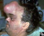 |
Neuro-Ophthalmic Manifestations of Brain Tumors | The patient is a 45-year-old recluse found to harbor this frontal lobe mass. Remarkably, this patient had only mild bilateral optic neuropathies with visual acuities in the 20/25 range. This right disc was mildly swollen and the left mildly pale. He could not fit into the fundus camera for disc phot... | Meningioma |
| 104 |
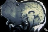 |
Neuro-Ophthalmic Manifestations of Brain Tumors | The patient is a 45-year-old recluse found to harbor this frontal lobe mass. Remarkably, this patient had only mild bilateral optic neuropathies with visual acuities in the 20/25 range. This right disc was mildly swollen and the left mildly pale. He could not fit into the fundus camera for disc phot... | Meningioma |
| 105 |
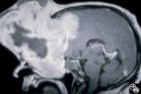 |
Neuro-Ophthalmic Manifestations of Brain Tumors | The patient is a 45-year-old recluse found to harbor this frontal lobe mass. Remarkably, this patient had only mild bilateral optic neuropathies with visual acuities in the 20/25 range. This right disc was mildly swollen and the left mildly pale. He could not fit into the fundus camera for disc phot... | Meningioma |
| 106 |
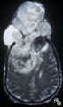 |
Neuro-Ophthalmic Manifestations of Brain Tumors | The patient is a 45-year-old recluse found to harbor this frontal lobe mass. Remarkably, this patient had only mild bilateral optic neuropathies with visual acuities in the 20/25 range. This right disc was mildly swollen and the left mildly pale. He could not fit into the fundus camera for disc phot... | Meningioma |
| 107 |
 |
Neuro-Ophthalmic Manifestations of Brain Tumors | The patient is a 45-year-old recluse found to harbor this frontal lobe mass. Remarkably, this patient had only mild bilateral optic neuropathies with visual acuities in the 20/25 range. This right disc was mildly swollen and the left mildly pale. He could not fit into the fundus camera for disc phot... | Meningioma |
| 108 |
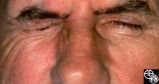 |
Ocular Manifestations of Systemic Disorders | This 59-year-old man had a 4-week history of malaise, myalgias, night sweats, weight loss, fatigue, and generalized headaches, and a few days before admission he developed a red eye OS secondary to exposure keratitis associated with a left-sided seventh nerve palsy. Subsequent evaluation disclosed a... | Lyme Disease |
| 109 |
 |
Ocular Manifestations of Congenital/Inherited Diseases | This 9-year-old girl, who had complained of recurrent spontaneous bleeding from the palate and slight swelling and increased warmth over the left cheek, was found to have Wyburn-Mason syndrome. Image 1993_16 shows a small area of arteriovenous shunt on the left optic disc in this patient, who has no... | Wyburn-Mason Syndrome |
| 110 |
 |
Ocular Manifestations of Congenital/Inherited Diseases | This 9-year-old girl, who had complained of recurrent spontaneous bleeding from the palate and slight swelling and increased warmth over the left cheek, was found to have Wyburn-Mason syndrome. Image 1993_16 shows a small area of arteriovenous shunt on the left optic disc in this patient, who has no... | Wyburn-Mason Syndrome |
| 111 |
 |
Ocular Manifestations of Congenital/Inherited Diseases | This 9-year-old girl, who had complained of recurrent spontaneous bleeding from the palate and slight swelling and increased warmth over the left cheek, was found to have Wyburn-Mason syndrome. Image 1993_16 shows a small area of arteriovenous shunt on the left optic disc in this patient, who has no... | Wyburn-Mason Syndrome |
| 112 |
 |
Ocular Manifestations of Congenital/Inherited Diseases | This 9-year-old girl, who had complained of recurrent spontaneous bleeding from the palate and slight swelling and increased warmth over the left cheek, was found to have Wyburn-Mason syndrome. Image 1993_16 shows a small area of arteriovenous shunt on the left optic disc in this patient, who has no... | Wyburn-Mason Syndrome |
| 113 |
 |
Ocular Manifestations of Congenital/Inherited Diseases | This 9-year-old girl, who had complained of recurrent spontaneous bleeding from the palate and slight swelling and increased warmth over the left cheek, was found to have Wyburn-Mason syndrome. Image 1993_16 shows a small area of arteriovenous shunt on the left optic disc in this patient, who has no... | Wyburn-Mason Syndrome |
| 114 |
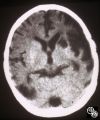 |
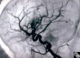
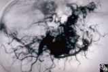
Neuro-Ophthalmic Vascular Disease | A 9-year-old boy had recurrent ischemic episodes that had begun 2 years prior to evaluation. A significant right hemiparesis and a significant speech, learning, and memory disorder were present. His noncontrast axial view CT scan demonstrated multiple cerebral infarcts. Cerebral angiography revealed... | Moyamoya Disease |
| 115 |
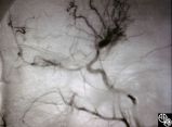 |
Neuro-Ophthalmic Vascular Disease | A 9-year-old boy had recurrent ischemic episodes that had begun 2 years prior to evaluation. A significant right hemiparesis and a significant speech, learning, and memory disorder were present. His noncontrast axial view CT scan demonstrated multiple cerebral infarcts. Cerebral angiography revealed... | Moyamoya Disease |
| 116 |
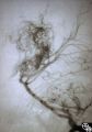 |
Neuro-Ophthalmic Vascular Disease | A 9-year-old boy had recurrent ischemic episodes that had begun 2 years prior to evaluation. A significant right hemiparesis and a significant speech, learning, and memory disorder were present. His noncontrast axial view CT scan demonstrated multiple cerebral infarcts. Cerebral angiography revealed... | Moyamoya Disease |
| 117 |
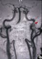 |
Neuro-Ophthalmic Vascular Disease | MR angiography was performed on this 33-year-old woman, who complained of the onset of a bad taste in her mouth followed by pain along the left forehead and development of the left third-order Horner's syndrome during pregnancy. Except for the Horner's syndrome, the patient was neurologically intact... | Carotid Dissection |
| 118 |
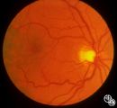 |
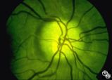
Neuro-Ophthalmic Vascular Disease | Image 93_28 shows the fundus before the attack. | Vasospastic Amaurosis Fugax |
| 119 |
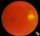 |
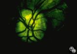
Neuro-Ophthalmic Vascular Disease | In image 93_29, taken during the episode, note the change in caliber of the blood vessels. | Vasospastic Amaurosis Fugax |
| 120 |
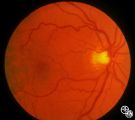 |
Neuro-Ophthalmic Vascular Disease | Image 93_30 is immediately after the attack, note the slight redness to the macula. | Vasospastic Amaurosis Fugax |
| 121 |
 |
Motility Disturbances | The patient is a 53-year-old man with diplopia from right oculomotor nerve palsy and left hemiparesis (Weber's syndrome), with associated left lung hilar mass. The spinal tap showed pleocytosis consistent with carcinomatous meningitis. This image demonstrates oculomotor nerve metastatic carcinomatos... | Oculomotor Palsy; Weber; Fascicular Oculomotor (Third) Nerve Palsy |
| 122 |
 |
Isolated Congenital Optic Disc Anomalies | This optic disc displays multiple drusen. Note the pseudopapilledema here. One can differentiate this from true papilledema in that there is no obscuration of the vessel by the peripapillary nerve fiber layer as they cross the disc margin. This photograph was taken with barrier filters in place, but... | Optic Disc Drusen; Optic Nerve Drusen; Pseudopapilledema |
| 123 |
 |
Optic Disc Drusen With Autofluorescence | This photograph of optic disc drusen demonstrates autoflourescence with flourescein barrier filters in place. Imaging: flourescein barrier filters. | Optic Disc Drusen; Autofluorescence |
| 124 |
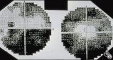 |
Optic Disc Drusen Visual Fields | This is the visual field of patient with optic nerve drusen. Whereas they typically do not cause central field loss, optic disc drusen may cause nerve fiber bundle layer defects and, thus, peripheral field defects, including altitudinal defects (seen inferiorly in the left eye) or arcuate defects (s... | Optic Disc Drusen; Optic Nerve Drusen; Visual Fields |
| 125 |
 |
Isolated Congenital Optic Disc Anomalies | Benign tumors of blood vessels (hemangiomas) may occur on the optic nerve and may mimic optic disc edema. Disease/Diagnosis: Optic Nerve Hemangioma. | Optic Nerve Hemangioma |