Best known for his world-renowned neuro-ophthalmology unit based at the University of California, San Francisco, William Hoyt, MD collected here more than 850 of his best images covering a wide range of disorders.
William F. Hoyt, MD, Professor Emeritus of Ophthalmology, Neurology and Neurosurgery, Department of Ophthalmology, University of California, San Francisco.
NOVEL: https://novel.utah.edu/
TO
Filters: Collection: "ehsl_novel_wfh"
| Title | Description | Type | ||
|---|---|---|---|---|
| 101 |
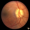 |
Vascular Complications of Drusen: Drusen Causing Loss of Superior Retinal Arterial Supply | PP32a: right; PP32b: left eye. Right eye is an obvious drusen disc. Patient had marked field defects. Left eye has occlusion of superior branch of the central retinal artery at 11:30 with the inferior retinal artery supplying collateral to the superior retina. Notice the branch of the inferior ret... | Image |
| 102 |
 |
Visible Drusen | PP24a. Right eye. Exposed drusen. There are inferior nerve fiber layer defects in the upper arcuate bundles. Optic disc is also hypoplastic. Anatomy: Optic disc. Pathology: Drusen of the optic disc. Disease/Diagnosis: Drusen of the optic disc. Clinical: Hypoplastic optic disc with drusen. | Image |
| 103 |
 |
Von Hippel Lindau Disease with Photocoagulation Effect | Von Hippel Lindau Disease with appearance of Xenon photocoagulation on a mini hemangioblastoma. Anatomy: Retina. Pathology: Hemangioblastoma. Disease/Diagnosis: Von Hippel Lindau disease. Clinical: No visual symptoms. | Image |
| 104 |
 |
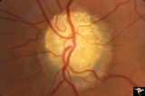
Unilateral Pseudopapilledema | PP_10a: Left: pseudo papilledema with disc blurring, crowded disc. Optic disc is small in diameter. PP_10b shows albinotic fundus and small crowded disc. Anatomy: Optic disc. Pathology: Normal variation of the optic disc. Disease/Diagnosis: Normal variation of the optic disc. Elevated disc. Clinic... | Image |
| 105 |
 |
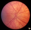
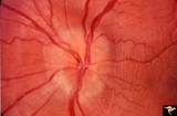
Bilateral Crowded Discs | Left eye. Bilateral crowded discs with congenital blurring. Blurred disc margins are not from edema. Note optic cup is absent. Pair with right eye in PP_1a, and brother in PP_2. Mother has drusen of the optic disc in PP_11aa & b. Sister has drusen in PP_11c. Anatomy: Optic disc. Pathology: Normal va... | Image |
| 106 |
 |
Buried Drusen | Buried drusen with peculiar white dot, which appears to be choroidal in location. Note lumpy disc margin on right disc PP_15a is right eye. PP_15b is left eye. Beautiful example of pseudo papilledema in which drusen can not be seen. 8 year old girl. Anatomy: Optic disc. Pathology: Drusen of the op... | Image |
| 107 |
 |
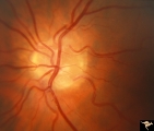
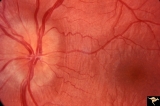
PP7a Crowded disc | PP7a: right eye crowded disc with blurred margin. Note anomalous vascular pattern and glial tissue on the disc; PP7b- left disc is cupless disc and normal. 10 year old girl with gonadal dysgenesis and growth retardation. Anatomy: Optic disc. Pathology: Normal variation of the optic disc. Disease/ Di... | Image |
| 108 |
 |
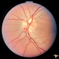
Unilateral Pseudopapilledema | PP_10b: shows albinotic fundus and a small crowded disc. PP_10a: left: pseudo papilledema with disc blurring, crowded disc. Optic disc is small in diameter. Anatomy: Optic disc. Pathology: Normal variation of the optic disc. Disease/Diagnosis: Normal variation of the optic disc. Crowded disc. Clin... | Image |
| 109 |
 |
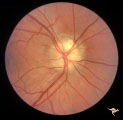
Vascular Complications of Drusen | PP34a: Right eye. Superior retinal vein drains into the choroid at 12:00. It has occluded between center of disc and 12:00. Note white ghost vessel. Note that other veins drain into the disc edge at 4:00. There is no evidence of a central retinal vein in the middle of the disc. PP34b: Visible drus... | Image |
| 110 |
 |
Vascular Complications of Drusen | PP34a: Right eye. Superior retinal vein drains into the choroid at 12:00. It has occluded between center of disc and 12:00. Note white ghost vessel. Note that other veins drain into the disc edge at 4:00. There is no evidence of a central retinal vein in the middle of the disc. PP34b: Visible drus... | Image |
| 111 |
 |
Vascular Complications of Drusen: Drusen Causing Loss of Superior Retinal Arterial Supply | PP32a: right; PP32b: left eye. Left eye has occlusion of superior branch of the central retinal artery at 11:30 with the inferior retinal artery supplying collateral to the superior retina. Notice the branch of the inferior retinal artery moves superiorly heading toward the upper retina. Drusen w... | Image |
| 112 |
 |
Visible Drusen with Visual Field Loss | Right eye visual field combine with PP25a, b, & d. Anatomy: Optic disc. Pathology: Drusen of the optic disc. Disease/Diagnosis: Drusen of the optic disc. Clinical: Drusen disc with severe visual field defect. note the nasal visual field loss and the arcuate bundle defects. Central vision was 20/20. | Image |
| 113 |
 |
Visible Drusen with Visual Field Loss | Left eye visual field. Combine with PP25 a, b, & c. Anatomy: Optic disc. Pathology: Drusen of the optic disc. Disease/Diagnosis: Drusen of the optic disc. Clinical: Note marked constriction of visual field in all quadrants with central preservation of vision. | Image |
| 114 |
 |
Visible Drusen with Visual Field Loss | PP25b right eye with drusen and severe visual field loss. Match with PP25a, c & d. Anatomy: Optic disc. Pathology: Drusen of the optic disc. Disease/Diagnosis: Drusen of the optic disc. Clinical: Drusen disc with servere visual field loss. | Image |
| 115 |
 |
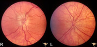
Visible Drusen - Bilateral | PP22a: right eye. PP22b: Note bypass vein draining into the choroid at 8:00. Anatomy: Optic disc. Pathology: Drusen of the optic disc. Disease/Diagnosis: Drusen of the optic disc. Clinical: Normally functioning eye with drusen. | Image |
| 116 |
 |
Buried Drusen with Sub-retinal Neovascular Net | Buried drusen with sub-retinal neovascular net. This is the same left eye. Appearance of the central retina of the left eye. Both PP29a & b are left eye: 17 year old girl; Visual acuity 10/400. Anatomy: Optic disc. Pathology: Drusen of the optic disc. DIsease/Diagnosis: Drusen of the optic disc. Cl... | Image |
| 117 |
 |
Drusen with Vertical Retinal Folds | PP36a & b: Both left eye: Buried drusen. Note vertical retinal folds. Anatomy: Optic disc. Pathology: Drusen of the optic disc. Disease/Diagnosis: Drusen of the optic disc. | Image |
| 118 |
 |
Drusen with Vertical Retinal Folds | PP36a & b:Both left eye: Buried drusen. Note vertical retinal folds. Anatomy: Optic disc. Pathology: Drusen of the optic disc. Disease/Diagnosis: Drusen of the optic disc. | Image |
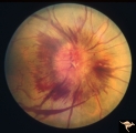
| 119 |
 |
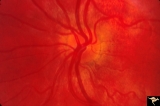
Bilateral Hemorrhagic Papilledema | Left eye. Bilateral Hemorrhagic Papilledema from cardio-respiratory disease. Woman. Anatomy: Optic disc. Pathology: Bilateral papilledema, hemorrhagic. Disease/Diagnosis: Pseudotumor due to cardio-respiratory disease. Clinical notes: Woman with headache, shortness of breath. | Image |
| 120 |
 |
Bilateral Hemorrhagic Papilledema | Bilateral Hemorrhagic Papilledema from cardio-respiratory disease. Woman. Anatomy: Optic disc. Pathology: Bilateral papilledema, hemorrhagic. Disease/Diagnosis: Pseudotumor due to cardio-respiratory disease. Clinical notes: Woman with headache, shortness of breath. | Image |
| 121 |
 |
Bilateral Papilledema | Right eye. Bilateral Papilledema in 410 pound man with tracheostomy for pulmonary insufficiency. Anatomy: Optic disc. Pathology: Papilledema. Disease/Diagnosis: Pseudotumor due to: sleep apnea due to cardiopulmonary insufficiency syndrome. Pickwickian syndrome. Clinical notes: Headache; obesity. | Image |
| 122 |
 |
Bilateral Papilledema | Picture of patient. 410 pound man with tracheostomy done for sleep apnea due to cardiopulmonary insufficiency syndrome. Pickwickian syndrome. Anatomy: Optic disc. Pathology: Papilledema. Disease/Diagnosis: Pseudotumor due to: sleep apnea due to cardiopulmonary insufficiency syndrome. Pickwickian syn... | Image |
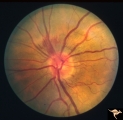
| 123 |
 |
Early Papilledema due to Tumor | Left eye. Asymmetric Papilledema with posterior fossa hemangioblastoma. Left - moderate papilledema. Blurring of disc. Young man. Anatomy: Optic disc. Pathology: Papilledema. Disease/Diagnosis: Papilledema from posterior fossa hemangioblastoma. | Image |
| 124 |
 |
P52b Asymmetric Papilledema with Choroidal Folds | Right eye shows papilledema. Asymmetric papilledema with choroidal folds. Bilateral choroidal folds from elevated intracranial pressure. Anatomy: Optic disc. Pathology: Papilledema. Disease/ Diagnosis: Chronic papilledema with choroidal folds. | Image |
| 125 |
 |
Post Papilledema with Choroidal Folds | Right eye. Post papilledema with choroidal folds due to brain tumor. Anatomy: Optic disc. Pathology: Post papilledema. Disease/Diagnosis: Post papilledema with choroidal folds. | Image |