Collection of materials relating to neuro-ophthalmology as part of the Neuro-Ophthalmology Virtual Education Library.
NOVEL: https://novel.utah.edu/
TO
- NOVEL981
Filters: Collection: "ehsl_novel_novel"
| Title | Creator | Description | Subject | ||
|---|---|---|---|---|---|
| 101 |
 |
Sports Related Head Injuries | Jessica Darusz, MD; Sean Gratton, MD | This narrated PowerPoint reviews the basics of sports related head injuries. It emphasizes assessment tools and treatment decisions in assessing athletes with concussion and other head injuries. | Concussion; Sports-related Head Injuries; Post-concussion Syndrome |
| 102 |
 |
Postconcussion Syndrome and Postconcussion Headache | Jessica Darusz, MD; Sean Gratton, MD | This brief presentation describes the pathophysiology, evaluation, and management of concussion, with an emphasis on postconcussion headache. | Concussion; Postconcussion Syndrome; Postconcussion Headache |
| 103 |
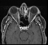 |
Curtain Sign (Enhanced Ptosis) - Associated Image 2 | Bashaer Aldhahwani, MD; Hong Jiang, MD, PhD | This is a 78-year-old male patient who presented with diplopia, right eyelid ptosis, and ophthalmoplegia. He had severe ptosis OD and pseudo-proptosis (lid retraction) OS at baseline, but when the right eyelid was manually elevated, there was marked enhanced ptosis of the left eyelid (Video). He was... | Myasthenia GravIs; Clinical Signs |
| 104 |
 |
Curtain Sign (Enhanced Ptosis) | Bashaer Aldhahwani, MD; Hong Jiang, MD, PhD | This is a 78-year-old male patient who presented with diplopia, right eyelid ptosis, and ophthalmoplegia. He had severe ptosis OD and pseudo-proptosis (lid retraction) OS at baseline, but when the right eyelid was manually elevated, there was marked enhanced ptosis of the left eyelid (Video). He was... | Myasthenia GravIs; Clinical Signs |
| 105 |
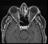 |
Curtain Sign (Enhanced Ptosis) - Associated Image 1 | Bashaer Aldhahwani, MD; Hong Jiang, MD, PhD | This is a 78-year-old male patient who presented with diplopia, right eyelid ptosis, and ophthalmoplegia. He had severe ptosis OD and pseudo-proptosis (lid retraction) OS at baseline, but when the right eyelid was manually elevated, there was marked enhanced ptosis of the left eyelid (Video). He was... | Myasthenia GravIs; Clinical Signs |
| 106 |
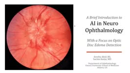 |
A Brief Introduction to AI in Neuro-ophthalmology | Areeba Abid, BS; Sachin Kedar, MD | In this video, we describe the basics of artificial intelligence and machine learning as applicable to clinical neuro-ophthalmologists. We use the example from a recent publication, where AI software was used to detect optic disc edema in fundus images. | Artificial Intelligence; Machine Learning |
| 107 |
 |
Morning Glory Disc Anomaly | Bashaer Aldhahwani, MD; Carlos Ernesto Mendoza Santiesteban, MD | A colored fundus photo of a patient with morning glory anomaly. Morning glory anomaly is a rare congenital malformation of the optic nerve. The morning glory disc anomaly can be seen with transsphenoidal basal encephalocele. It is known as morning glory syndrome when it is associated with systemic s... | Optic Disc Anomaly |
| 108 |
 |
Headaches Related to Eye Disorders | Nikki Gill, MSIII; Sean Gratton, MD | Headache attributed to disorders of the eye: Angle closure glaucoma, Ocular inflammation, Refractive error, Trochlear headache, Ocular surface disorder | Eye Disorders; Headache; Angle Closure Glaucoma; Ocular Inflammation; Refractive Error; Trochlear Headache; Ocular Surface Disorder |
| 109 |
 |
Introduction to NANOS NOTE | Karl C. Golnik, MD | Introduction to NANOS NOTE, a resource for non-neuro-ophthalmologists describing common examination techniques. | Neuro-Ophthalmology Examination Techniques |
| 110 |
 |
Optic Nerve Hypoplasia (ONH) - Double Ring Sign | Bashaer Aldhahwani, MD; Joshua Pasol, MD | Optic nerve hypoplasia (ONH) is characterized by a decreased number of optic nerve axons. It can present unilaterally or bilaterally, Isolated or associated with midline cerebral structural defects, such as septum pellucidum absence, agenesis of corpus callosum, cerebral hemisphere abnormalities, or... | Optic Nerve Hypoplasia (ONH) |
| 111 |
 |
Myelinated Retinal Nerve Fiber Layer (MRNFL) | Sparsh Jain, BS; Ryan D. Walsh, MD | Fundus photos demonstrating bilateral (right > left) peripapillary myelinated retinal nerve fiber layer (MRNFL) in a 14-year old boy. Note the typical appearance of MRNFL of a white patch with feathered margins involving the inner retina. In this case, the MRNFL is more prominent in the right eye, a... | Myelinated Retinal Nerve Fiber Layer; MRNFL; Congenital Anomalies |
| 112 |
 |
Vitamin B12 Deficiency and Neuro-Ophthalmic Manifestations | Jourdan Carroll; Devin D. Mackay, MD | This presentation covers vitamin B12 deficiency, including etiology, signs and symptoms, neurologic and ophthalmic findings, a case presentation and treatment. | Vitamin B12 Deficiency and Neuro-Ophthalmic Manifestations |
| 113 |
 |
Ocular Neuromyotonia Video | Bashaer Aldhahwani, MD; Joshua Pasol, MD | A video demonstrates ocular neuromyotonia in the left eye of a patient with a history of cranial radiation of parasellar mass. Ocular neuromyotonia (ONM) is a rare ocular motor disorder characterized by intermittent, tonic spasms of one or more of the extraocular muscles, resulting in strabismus and... | Ocular Neuromyotonia |
| 114 |
 |
Myelinated Retinal Nerve Fiber Layer | Bashaer Aldhahwani, MD; Hong Jiang, MD, PhD | A 78 YOF with no visual symptoms has an incidental finding of yellow-white well-demarcated patches with ragged borders at the peripapillary area of her left eye (see the fundus photo). | Myelinated Retinal Nerve Fiber Layer |
| 115 |
 |
Cogan Lid Twitch | Hari Anandarajah, BA | A 50-year-old woman presented with ptosis of her left eyelid for 6 months. Several exam findings including variable and fatigable ptosis, and Cogan lid twitch, raised suspicion for Myasthenia Gravis. Acetylcholine receptor binding, blocking, and modulating antibodies were negative, and single fiber ... | Lid Twitch; Myasthenia Gravis |
| 116 |
 |
Behcet's Disease | Harinee Arunachalam, MSIV; Sean Gratton, MD | This video provides an overview of Behcet's Disease, a rare vasculitis of unknown etiology. | Behcet's Disease |
| 117 |
 |
Indirect Carotid Cavernous Fistula | Edsel Ing, MD, PhD, FRCSC | A 67-year-old woman had delayed initial diagnosis of her right low flow carotid cavernous fistula (CCF) during the coronavirus disease (COVID-19) pandemic due to difficulty detecting ocular signs via online virtual examinations. Her right eye conjunctival erythema and proptosis with medial rectus en... | Carotid Cavernous Fistula; Misdiagnosis; Radiology |
| 118 |
 |
Giant Cell Arteritis: Diagnostic Prediction Models, Temporal Artery Biopsy and Epidemiology | Edsel Ing MD, PhD FRCSC MPH CPH MIAD MEd MBA, | Giant cell arteritis (GCA) is the most common primary vasculitis in the elderly and can cause irreversible blindness, aortitis, and stroke. Diagnostic confirmation of GCA usually entails temporal artery biopsy (TABx) - a time-consuming and invasive test, or ultrasound. The primary treatment of GCA i... | Giant Cell Arteritis; Diagnostic Prediction Model; Epidemiology; Temporal Artery Biopsy; Differential Diagnosis |
| 119 |
 |
Thrombectomy for Acute Ischemic Stroke | Aaron W. Grossman, MD, PhD | A video overview of thrombectomy of acute ischemic stroke. Covers anatomy, history of treatment, and current practice. | Thrombectomy; Acute Ischemic Stroke |
| 120 |
 |
Direct-Indirect Ophthalmoscopy (DIO) | Irina Krikova, PA-C; Eric Caskey, MD; Alison Crum, MD; Kathleen Digre, MD; James Gilman, CRA, FOPS; Levi Goldfarb, MBA, MD Candidate; Bradley Katz, MD; Ethan Peterson, Videographer; Meagan Seay, DO; Judith Warner, MD | A slideshow describing the use of the direct ophthalmoscope. | Ophthalmoscopy |
| 121 |
 |
Vogt-Koyanagi-Harada Syndrome | Shwetha Mudalegundi, Medical Student; Amanda D. Henderson, MD | Vogt-Koyanagi-Harada (VKH) syndrome is a rare disorder that affects several body systems. Here we take a broad look at the presentation and pathophysiology of VKH, with a more specific focus on the relevant eye findings. Since much is not known about VKH, we explore the current standards for diagnos... | Vogt-Koyanagi-Harada Syndrome; Uveitis |
| 122 |
 |
Surgical Management of Strabismus | Michelle S. Attzs, MBBS, FRCOphth | This is a brief introduction to the surgical management of strabismus. It includes the key elements of the work up for a patient about to undergo strabismus surgery, introduces the basics on surgical techniques including adjustable sutures, and discusses the complications associated with this surger... | Strabismus; Surgery; Ocular Motility; Adjustable Sutures; Esotropia; Exotropia; Complications |
| 123 |
 |
A Case Series of Mydriasis from an Anticholinergic Antiperspirant | Aileen Antonio, MD; Inna Bondira, DO; Cameron Holicki, DO; Christopher Glisson, DO; Tatiana Deveney, MD; Lina Nagia, DO | Causes of anisocoria span a wide range, from benign to life-threatening, making it a common indication for Neuro-Ophthalmology referrals. One such cause is related to pharmacologic mydriasis due to direct or systemic exposure. We present a case series of four patients with different presentations of... | Anisocoria; Mydriasis; Pharmacologic Anisocoria; Anticholinergic Antiperspirant |
| 124 |
 |
Radiation Optic Neuropathy | Khawla Elnour; Amanda Henderson, MD | A video describing optic neuropathy related to radiation. | Radiation; Neuropathy |
| 125 |
 |
Optical Coherence Tomography (OCT) | Gunnar J. Goebel; Devin D. Mackay, MD | Introduction to OCT, including history, principles, interpretation, and applications. | Optical Coherence Tomography (OCT) |