AAO-NANOS Neuro-Ophthalmology Clinical Collection: Derived from the AAO-NANOS Clinical Neuro-Ophthalmology collection produced on CD. The images are of selected cases from the NANOS teaching slide exchange, and the CD was produced under the direction of Larry Frohman, MD and Andrew Lee, MD.
The American Academy of Ophthalmology (AAO); The North American Neuro-Ophthalmology Association (NANOS).
NOVEL: https://novel.utah.edu/
TO
Filters: Collection: "ehsl_novel_aao_nanos"
| Title | Creator | Description | ||
|---|---|---|---|---|
| 101 |
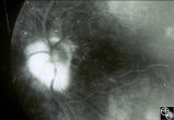 |
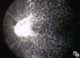
Isolated Congenital Optic Disc Anomalies | Larry P. Frohman, MD | This 63-year-old man with amblyopia OD was seen for a question of ischemic optic neuropathy with a pale, swollen disc OD. The correct diagnosis is an exophytic capillary angioma of the optic nerve head. Disease/Diagnosis: Capillary Angioma. |
| 102 |
 |
Optic Neuropathies | Larry P. Frohman, MD | This healthy 29-year-old man with dense amblyopia OS presented with a foreign-body sensation OS and further visual loss in his amblyopic eye. He was noted to have bilateral disc edema and lesions in the left eye consistent with unilateral acute multifocal placoid pigment epitheliopathy (AMPPE). He r... |
| 103 |
 |
Optic Neuropathies | Larry P. Frohman, MD | This healthy 29-year-old man with dense amblyopia OS presented with a foreign-body sensation OS and further visual loss in his amblyopic eye. He was noted to have bilateral disc edema and lesions in the left eye consistent with unilateral acute multifocal placoid pigment epitheliopathy (AMPPE). He r... |
| 104 |
 |
Optic Neuropathies | Larry P. Frohman, MD | This healthy 29-year-old man with dense amblyopia OS presented with a foreign-body sensation OS and further visual loss in his amblyopic eye. He was noted to have bilateral disc edema and lesions in the left eye consistent with unilateral acute multifocal placoid pigment epitheliopathy (AMPPE). He r... |
| 105 |
 |
Optic Neuropathies | Larry P. Frohman, MD | This healthy 29-year-old man with dense amblyopia OS presented with a foreign-body sensation OS and further visual loss in his amblyopic eye. He was noted to have bilateral disc edema and lesions in the left eye consistent with unilateral acute multifocal placoid pigment epitheliopathy (AMPPE). He r... |
| 106 |
 |
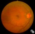
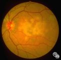
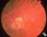
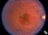
Systemic Disorders With Optic Nerve and Retinal Findings | Larry P. Frohman, MD | At age 41, in 1984, this woman, who grew up in the Ohio River Valley, had 3 days of ocular pain OD, and her vision declined to 20/80 OD she has had no visual changes since, nor has she had any other neurologic symptoms. The ""presumed"" diagnosis is optic neuropathy in presumed ocular histoplasmosis... |
| 107 |
 |
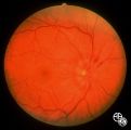
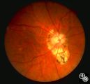
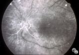
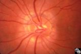
Systemic Disorders With Optic Nerve and Retinal Findings | Larry P. Frohman, MD | This 57-year-old man had a neuro-ophthalmology consult, requested the night before his 2-cm pituitary tumor was to be resected. His examination revealed his acuities to be 20/70 OU, with a visual field not consistent with chiasmal compression. The fundus appearance, with peripheral salt and pepperin... |
| 108 |
 |
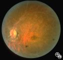
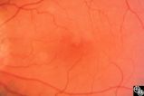
Neuro-Ophthalmic Vascular Disease | Steven A. Newman, MD | Occlusion of a branch or central retinal artery may result in acute visual loss. The ophthalmoscopic findings are retinal whitening due to ischemic retina in the distribution of the occluded artery. Sparing or selective involvement of cilioretinal artery branches may occur. Patients with a central r... |
| 109 |
 |
Neuro-Ophthalmic Vascular Disease | Steven A. Newman, MD | Occlusion of a branch or central retinal artery may result in acute visual loss. The ophthalmoscopic findings are retinal whitening due to ischemic retina in the distribution of the occluded artery. Sparing or selective involvement of cilioretinal artery branches may occur. Patients with a central r... |
| 110 |
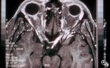 |
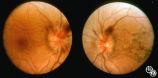
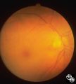
Neuro-Ophthalmic Imaging-MRI | Scott Forman, MD | This 23-year-old right-handed man had a history of idiopathic recurrent optic neuritis. The patient presented with acuity of 20/400 OD and 20/100 OS, with a central scotoma OD and a complete temporal defect OS. MRI with fat suppression and gadolinium revealed enhancement of the intracranial nerve an... |
| 111 |
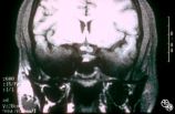 |
Neuro-Ophthalmic Imaging-MRI | Scott Forman, MD | This 23-year-old right-handed man had a history of idiopathic recurrent optic neuritis. The patient presented with acuity of 20/400 OD and 20/100 OS, with a central scotoma OD and a complete temporal defect OS. MRI with fat suppression and gadolinium revealed enhancement of the intracranial nerve an... |
| 112 |
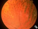 |
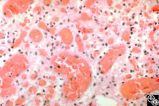
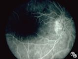
Systemic Disorders With Optic Nerve and Retinal Findings | Robert L. Lesser, MD | Intraocular lymphoma may present with an unexplained vitritis, optic disc infiltration, or choroidal infiltration. One unusual manifestation of large-cell lymphoma is this leopard-spot appearance. Pair with 94_32, 94_33, and 94_35. This is a fundus photo. |
| 113 |
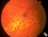 |
Systemic Disorders With Optic Nerve and Retinal Findings | Robert L. Lesser, MD | Intraocular lymphoma may present with an unexplained vitritis, optic disc infiltration, or choroidal infiltration. One unusual manifestation of large-cell lymphoma is this leopard-spot appearance. Pair with 94_32, 94_34, and 94_35. This is a fundus photo. |
| 114 |
 |
Systemic Disorders With Optic Nerve and Retinal Findings | Robert L. Lesser, MD | Intraocular lymphoma may present with an unexplained vitritis, optic disc infiltration, or choroidal infiltration. One unusual manifestation of large-cell lymphoma is this leopard-spot appearance. Pair with 94_33, 94_34, and 94_35. This is a fundus photo. |
| 115 |
 |
Optic Neuropathies | Daniel M. Jacobson MD | This 28-year-old otherwise-healthy woman was referred to for treatment of what was thought to be optic neuritis OD. Three weeks earlier she had noted a dark inferior scotoma OD that progressed to involve fixation over the next 10-12 days. She experienced photopsias OD at the onset. She had no viral ... |
| 116 |
 |
Optic Neuropathies | Daniel M. Jacobson MD | This 28-year-old otherwise-healthy woman was referred to for treatment of what was thought to be optic neuritis OD. Three weeks earlier she had noted a dark inferior scotoma OD that progressed to involve fixation over the next 10-12 days. She experienced photopsias OD at the onset. She had no viral ... |
| 117 |
 |
Optic Neuropathies | Daniel M. Jacobson MD | This 34-year-old otherwise-healthy woman was referred for a neuro-ophthalmologic consultation for unexplained loss of vision OD following head trauma. Two months earlier, she was involved in a motor vehicle accident that resulted in a closed head injury complicated by a spontaneously resolving small... |
| 118 |
 |
Neuro-Ophthalmic Vascular Disease | Larry P. Frohman, MD | This 27-year-old woman had no past ocular history and presented with 3 weeks of redness OS that has been treated by the referring doctor as allergic conjunctivitis. She was referred for evaluation when she developed binocular diplopia. Her past medical history included phlebitis and one miscarriage ... |
| 119 |
 |
Neuro-Ophthalmic Vascular Disease | Larry P. Frohman, MD | This 27-year-old woman had no past ocular history and presented with 3 weeks of redness OS that has been treated by the referring doctor as allergic conjunctivitis. She was referred for evaluation when she developed binocular diplopia. Her past medical history included phlebitis and one miscarriage ... |
| 120 |
 |
Ocular Manifestations of Congenital/Inherited Diseases | Steven Galetta, MD | This 21-year-old woman had a 2-year history of blurred vision. A computerized visual field demonstrated a temporal defect OS. MRI confirmed a chiasmal mass lesion. The pathology was consistent with hemangioblastoma. Further workup revealed retinal angiomas and multiple other hemangioblastomas of the... |
| 121 |
 |
Neuro-Ophthalmic Vascular Disease | Steven A. Newman, MD | Occlusion of a branch or central retinal artery may result in acute visual loss. The ophthalmoscopic findings are retinal whitening due to ischemic retina in the distribution of the occluded artery. Sparing or selective involvement of cilioretinal artery branches may occur. Patients with a central r... |
| 122 |
 |
Neuro-Ophthalmic Consequences of Therapy | Larry P. Frohman, MD | This woman presented at age 52, 3 years after radiation therapy for a salivary gland carcinoma extending into the right maxillary sinus. She had received 6000 rads in 30 fractions over 45 days. She presented with 3 weeks of visual loss, with acuity of 20/30, normal color plates, normal fields, and n... |
| 123 |
 |
Neuro-Ophthalmic Consequences of Therapy | Larry P. Frohman, MD | This woman presented at age 52, 3 years after radiation therapy for a salivary gland carcinoma extending into the right maxillary sinus. She had received 6000 rads in 30 fractions over 45 days. She presented with 3 weeks of visual loss, with acuity of 20/30, normal color plates, normal fields, and n... |
| 124 |
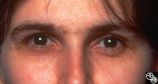 |
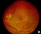
Systemic Disorders With Optic Nerve and Retinal Findings | Robert F. Saul, MD | This patent has known pseudoxanthoma elasticum (an uncommon elastic tissue disorder characterized by plaque-like skin folds [plucked chicken skin], and degeneration of collagen fibers involving multiple systems, including the GI tract and heart), angioid streaks, and optic disc drusen. |
| 125 |
 |
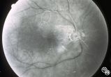
Systemic Disorders With Optic Nerve and Retinal Findings | Robert L. Lesser, MD | Intraocular lymphoma may present with an unexplained vitritis, optic disc infiltration, or choroidal infiltration. One unusual manifestation of large-cell lymphoma is this leopard-spot appearance. Pair with 94_32, 94_33, and 94_34. This is a fluorescein angiogram. |