Best known for his world-renowned neuro-ophthalmology unit based at the University of California, San Francisco, William Hoyt, MD collected here more than 850 of his best images covering a wide range of disorders.
William F. Hoyt, MD, Professor Emeritus of Ophthalmology, Neurology and Neurosurgery, Department of Ophthalmology, University of California, San Francisco.
NOVEL: https://novel.utah.edu/
TO
| Title | Description | Type | ||
|---|---|---|---|---|
| 101 |
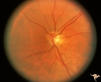 |
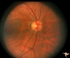
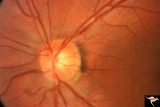
IF203b Temporal Cupping with Dominant Hereditary Optic Atrophy | Left disc is hypoplastic and smaller with temporal pallor. Pair with IF2_3a. 1994. Anatomy: Optic disc. Pathology: Dominant hereditary optic atrophy. Disease/ Diagnosis: Dominant hereditary optic atrophy. Clinical: Depressed central vision. | Image |
| 102 |
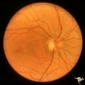 |
IF204a Temporal Cupping with Dominant Hereditary Optic Atrophy | Right eye with temporal pallor and shallow cupping. Pair with IF2_4b. 1960. Anatomy: Optic disc. Pathology: Dominant hereditary optic atrophy. Disease/ Diagnosis: Dominant hereditary optic atrophy. Clinical: Dominant hereditary optic atrophy. | Image |
| 103 |
 |
IF204b Temporal Cupping with Dominant Hereditary Optic Atrophy | Left eye with temporal pallor and shallow cupping. Pair with IF2_4a. 1960. Anatomy: Optic disc. Pathology: Dominant hereditary optic atrophy. Disease/ Diagnosis: Dominant hereditary optic atrophy. Clinical: Depressed central vision. | Image |
| 104 |
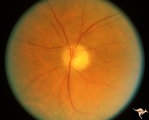 |
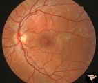
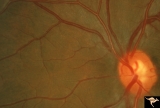
IF301 Post Giant Cell Arteritis Ischemic Papillopthy | Post giant cell arteritis ischemic papillopathy. Note shallow cupping without vascular displacement.1994. Also note the irregular focal arteriolar narrowing. Blind eye. Male. Anatomy: Optic disc. Pathology: Ischemic papillopathy from giant cell arteritis. Disease/ Diagnosis; Ischemic papillopathy fr... | Image |
| 105 |
 |
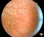
IIA101 Diffuse Atrophy | Primary or retrograde optic atrophy. It occurs from any injury to an optic nerve in the orbit or within the skull. Diffuse optic atrophy and blindness from nerve compression by pituitary tumor. Black woman. 1974. Note that retrograde optic atrophy can be associated with narrowed retinal arterioles i... | Image |
| 106 |
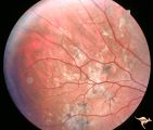 |
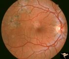
Multifocal Choroidopathy | Multifocal choroidopathy in a patient with uveitis. Anatomy: Retina. Disease/Diagnosis: Acute Multifocal Placoid Pigment Epitheliopathy (AMPPE). Clinical: Visual loss. | Image |
| 107 |
 |
Multifocal Choroidopathy | Multifocal choroidopathy in a patient with uveitis. Anatomy: Retina. Disease/Diagnosis: Acute Multifocal Placoid Pigment Epitheliopathy (AMPPE). Clinical: Visual loss. | Image |
| 108 |
 |
Multifocal Choroidopathy | Multifocal choroidopathy in a patient with uveitis. Anatomy: Retina. Disease/Diagnosis: Acute Multifocal Placoid Pigment Epitheliopathy (AMPPE). Clinical: Visual loss. | Image |
| 109 |
 |
Multifocal Choroidopathy | Multifocal choroidopathy in a patient with uveitis. Anatomy: Retina. Disease/Diagnosis: Acute Multifocal Placoid Pigment Epitheliopathy (AMPPE). Clinical: Visual loss. | Image |
| 110 |
 |
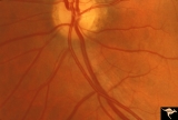
Multiple Sclerosis Slits and Thinning in Peripapillary (Retinal) Nerve Riber Layer | Multiple slit defect in the superior arcuate nerve fiber layer. Anatomy: Peripapillary nerve fiber layer. Pathology: Slit-like atrophy. Disease/Diagnosis: Multiple sclerosis optic neuropathy. Clinical: No symptoms. | Image |
| 111 |
 |
Multiple Sclerosis Slits and Thinning in Peripapillary (Retinal) Nerve Riber Layer | Multiple slit defect in the superior arcuate nerve fiber layer. Pair with IIB2_6b. Anatomy: Peripapillary nerve fiber layer. Pathology: Slit-like atrophy. Disease/Diagnosis: Multiple sclerosis optic neuropathy. Clinical: No symptoms. | Image |
| 112 |
 |
Multiple Sclerosis Slits and Thinning in Peripapillary (Retinal) Nerve Riber Layer | Multiple slit like defects in the inferior arcuate nerve fibers. Pair with IIB2_3b. Anatomy: Peripapillary nerve fiber layer. Pathology: Slit-like atrophy. Disease/Diagnosis: Multiple sclerosis optic neuropathy. Clinical: No symptoms. | Image |
| 113 |
 |
Multiple Sclerosis Slits and Thinning in Peripapillary (Retinal) Nerve Riber Layer | Multiple slit and wedge defects in the nerve fiber layer. Pair with IIB2_3a. Anatomy: Peripapillary nerve fiber layer. Pathology: Slit-like atrophy. Disease/Diagnosis: Multiple sclerosis optic neuropathy. Clinical: No symptoms. | Image |
| 114 |
 |
Multiple Sclerosis Slits and Thinning in Peripapillary (Retinal) Nerve Riber Layer | Multiple slit defect in the superior arcuate nerve fiber layer. Anatomy: Peripapillary nerve fiber layer. Pathology: Slit-like atrophy. Disease/Diagnosis: Multiple sclerosis optic neuropathy. Clinical: No symptoms. | Image |
| 115 |
 |
Multiple Sclerosis Slits and Thinning in Peripapillary (Retinal) Nerve Riber Layer | Multiple slit defect in the superior arcuate nerve fiber layer. Magnified. Pair with IIB2_6a. Anatomy: Peripapillary nerve fiber layer. Pathology: Slit-like atrophy. Disease/Diagnosis: Sclerosis optic neuropathy. Clinical: No symptoms. | Image |
| 116 |
 |
Multiple Sclerosis Slits and Thinning in Peripapillary (Retinal) Nerve Riber Layer | Left eye. Upper arcuate nerve fiber layer contains multiple low density slits. These indicate nerve fiber loss. Anatomy: Peripapillary nerve fiber layer. Pathology: Slit-like atrophy. Disease/Diagnosis: Multiple sclerosis optic neuropathy. Clinical: No symptoms. | Image |
| 117 |
 |
Multiple Sclerosis Slits and Thinning in Peripapillary (Retinal) Nerve Riber Layer | Multiple slit defect in the superior arcuate nerve fiber layer in a 13 year old boy. Right eye. Pair with IIB2_7a. Anatomy: Peripapillary nerve fiber layer. Pathology: Slit-like atrophy. Disease/Diagnosis: Multiple sclerosis optic neuropathy. Clinical: No symptoms. | Image |
| 118 |
 |
Multiple Sclerosis Slits and Thinning in Peripapillary (Retinal) Nerve Riber Layer | Multiple slit defect in the superior arcuate nerve fiber layer in a 13 year old boy. Pair with IIB2_7b. Anatomy: Peripapillary nerve fiber layer. Pathology: Slit-like atrophy. Disease/Diagnosis: Multiple sclerosis optic neuropathy. Clinical: No symptoms. | Image |
| 119 |
 |
Multiple Sclerosis Slits and Thinning in Peripapillary (Retinal) Nerve Riber Layer | Need magnification - Left eye - Peculiar punctate dotted surface of internal limiting membrane reflexes. Pairs with IIB2_01a & IIB2_02b. Anatomy: Peripapillary nerve fiber layer. Pathology: Slit-like atrophy. Disease/Diagnosis: Multiple sclerosis optic neuropathy. Clinical: No symptoms. | Image |
| 120 |
 |
Multiple Sclerosis Slits and Thinning in Peripapillary (Retinal) Nerve Riber Layer | Need magnification - Left eye - Inferior arcuate nerve fiber slits. Pairs with IIB2_01b & IIB2_01c. Anatomy: Peripapillary nerve fiber layer. Pathology: Slit-like atrophy. Disease/Diagnosis: Multiple sclerosis optic neuropathy. Clinical: No symptoms. | Image |
| 121 |
 |
Multiple Sclerosis Slits and Thinning in Peripapillary (Retinal) Nerve Riber Layer | Need magnification - Left eye - Inferior arcuate nerve fiber slits. Pairs with IIB2_01a & IIB2_01c. Anatomy: Peripapillary nerve fiber layer. Pathology: Slit-like atrophy. Disease/Diagnosis: Multiple sclerosis optic neuropathy. Clinical: No symptoms. | Image |
| 122 |
 |
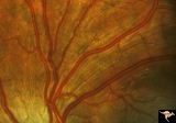
Normal Peripapillary Nerve Fiber Layer | Normal nerve fiber layer with Gunn's Dots visible in the upper arcuate fiber zone. This is a normal peripapillary nerve fiber layer in a young woman. Note the way the nerve fiber striations obscure and partially bury the small vessels running across them. Also note the interrupted surface reflex on ... | Image |
| 123 |
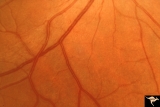 |
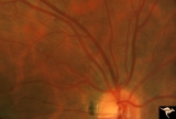
Normal Peripapillary Nerve Fiber Layer | Example of 23 year old woman, healthy nerve fiber layer below her optic disc. This is a normal peripapillary nerve fiber layer in a young woman. Note the way the nerve fiber striations obscure and partially bury the small vessels running across them. Also note the interrupted surface reflex on arter... | Image |
| 124 |
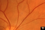 |
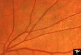
Normal Peripapillary Nerve Fiber Layer | Example of 23 year old woman, healthy nerve fiber layer below her optic disc. This is a normal peripapillary nerve fiber layer in a young woman. Note the way the nerve fiber striations obscure and partially bury the small vessels running across them. Also note the interrupted surface reflex on arter... | Image |
| 125 |
 |
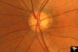
Normal Peripapillary Nerve Fiber Layer | This is a normal peripapillary nerve fiber layer in a young woman. Note the way the nerve fiber striations obscure and partially bury the small vessels running across them. Also note the interrupted surface reflex on arteries due to interposed nerve fiber layer tissue. All these vessels are buried i... | Image |