AAO-NANOS Neuro-Ophthalmology Clinical Collection: Derived from the AAO-NANOS Clinical Neuro-Ophthalmology collection produced on CD. The images are of selected cases from the NANOS teaching slide exchange, and the CD was produced under the direction of Larry Frohman, MD and Andrew Lee, MD.
The American Academy of Ophthalmology (AAO); The North American Neuro-Ophthalmology Association (NANOS).
NOVEL: https://novel.utah.edu/
TO
| Title | Creator | Description | ||
|---|---|---|---|---|
| 101 |
 |
Motility Disturbances | Larry P. Frohman, MD | This 5-year-old child presented with a 70 PD exotropia OS and a right face turn. She had a CT scan of the head at age 4 months that was normal , and she was felt to have an isolated left medial rectus paresis. Her acuity was 20/20 OU. She could fuse with a large face turn, and was orthomorphic is ex... |
| 102 |
 |
Motility Disturbances | Larry P. Frohman, MD | This 5-year-old child presented with a 70 PD exotropia OS and a right face turn. She had a CT scan of the head at age 4 months that was normal , and she was felt to have an isolated left medial rectus paresis. Her acuity was 20/20 OU. She could fuse with a large face turn, and was orthomorphic is ex... |
| 103 |
 |
Motility Disturbances | Larry P. Frohman, MD | This 5-year-old child presented with a 70 PD exotropia OS and a right face turn. She had a CT scan of the head at age 4 months that was normal , and she was felt to have an isolated left medial rectus paresis. Her acuity was 20/20 OU. She could fuse with a large face turn, and was orthomorphic is ex... |
| 104 |
 |
Systemic Disorders With Optic Nerve and Retinal Findings | Larry P. Frohman, MD | A 29-year-old African American woman presented with headaches, bilateral transient visual obscurations, blurred vision, numbness, and weakness of the lower extremities with myalgia and joint pains. She had an unplanned 12-pound weight loss over 2 months. A neurologist and internist diagnosed her wit... |
| 105 |
 |
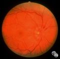
Systemic Disorders With Optic Nerve and Retinal Findings | Larry P. Frohman, MD | A 25-year-old African-American woman presented with a 20/200 optic neuropathology and no other illness. Because this disc appearance of a granuloma led us to suspect occult sarcoidosis, she underwent systemic evaluation. She was ultimately shown to have systemic sarcoidosis, including pulmonary invo... |
| 106 |
 |
Ocular Manifestations of Systemic Disorders | Larry P. Frohman, MD | A 17-year-old girl had undergone multiple superficial biopsies of the orbit for what was felt to be refractory orbital pseudotumor. Initial evaluation revealed the saddle-nose deformity, which the patient confirmed was acquired. More extensive biopsy was consistent with lymphomatoid granulomatosis. ... |
| 107 |
 |
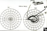
Optic Tract Syndrome Due to Carotid Artery Dolichoectasia | Larry P. Frohman, MD | This 43-year-old man was referred for evaluation of 6 months of visual loss OU. In retrospect, he had noticed increasing difficulty with his tennis game dating back over 3 years, as balls would pass him unexpectedly when hit to his backhand (left) side. The patient did not think this was progressive... |
| 108 |
 |
Optic Tract Syndrome Due to Carotid Artery Dolichoectasia | Larry P. Frohman, MD | This 43-year-old man was referred for evaluation of 6 months of visual loss OU. In retrospect, he had noticed increasing difficulty with his tennis game dating back over 3 years, as balls would pass him unexpectedly when hit to his backhand (left) side. The patient did not think this was progressive... |
| 109 |
 |
Isolated Optic Neuritis/Neuropathy | Larry P. Frohman, MD | The patient is a 62-year-old female who presented in August 1996 with visual loss OD that she first noted as loss of her superior field in May 1996. She felt that it had been static since, and perhaps was even a little better in the week before she was seen. There was no pain, even with ocular rotat... |
| 110 |
 |
Isolated Optic Neuritis/Neuropathy | Larry P. Frohman, MD | The patient is a 62-year-old female who presented in August 1996 with visual loss OD that she first noted as loss of her superior field in May 1996. She felt that it had been static since, and perhaps was even a little better in the week before she was seen. There was no pain, even with ocular rotat... |
| 111 |
 |
Isolated Optic Neuritis/Neuropathy | Larry P. Frohman, MD | The patient is a 62-year-old female who presented in August 1996 with visual loss OD that she first noted as loss of her superior field in May 1996. She felt that it had been static since, and perhaps was even a little better in the week before she was seen. There was no pain, even with ocular rotat... |
| 112 |
 |
Isolated Optic Neuritis/Neuropathy | Larry P. Frohman, MD | The patient is a 62-year-old female who presented in August 1996 with visual loss OD that she first noted as loss of her superior field in May 1996. She felt that it had been static since, and perhaps was even a little better in the week before she was seen. There was no pain, even with ocular rotat... |
| 113 |
 |
Isolated Optic Neuritis/Neuropathy | Larry P. Frohman, MD | The patient is a 62-year-old female who presented in August 1996 with visual loss OD that she first noted as loss of her superior field in May 1996. She felt that it had been static since, and perhaps was even a little better in the week before she was seen. There was no pain, even with ocular rotat... |
| 114 |
 |
Isolated Optic Neuritis/Neuropathy | Larry P. Frohman, MD | The patient is a 62-year-old female who presented in August 1996 with visual loss OD that she first noted as loss of her superior field in May 1996. She felt that it had been static since, and perhaps was even a little better in the week before she was seen. There was no pain, even with ocular rotat... |
| 115 |
 |
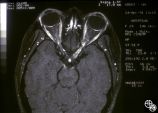
Orbital Tumors | Larry P. Frohman, MD | This 30-year-old man had a retrobulbar intraconal mass OS. The CT scans showed a heterogeneous lobulated enhancing mass, 2.2 x 1.9 x 1.8 cm. The case beautifully exhibits chorodial folds. The ultrasound showed internal reflectivity. The patient refused surgery. Pair with Images 97_60, 97_62, 97_63, ... |
| 116 |
 |
Orbital Tumors | Larry P. Frohman, MD | This 30-year-old man had a retrobulbar intraconal mass OS. The CT scans showed a heterogeneous lobulated enhancing mass, 2.2 x 1.9 x 1.8 cm. The case beautifully exhibits chorodial folds. The ultrasound showed internal reflectivity. The patient refused surgery. Pair with Images 97_60, 97_61, 97_63, ... |
| 117 |
 |
Orbital Tumors | Larry P. Frohman, MD | This 30-year-old man had a retrobulbar intraconal mass OS. The CT scans showed a heterogeneous lobulated enhancing mass, 2.2 x 1.9 x 1.8 cm. The case beautifully exhibits chorodial folds. The ultrasound showed internal reflectivity. The patient refused surgery. Pair with Images 97_61, 97_62, 97_63, ... |
| 118 |
 |
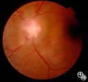
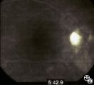
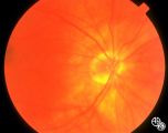
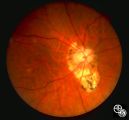
Isolated Congenital Optic Disc Anomalies | Larry P. Frohman, MD | This 63-year-old man with amblyopia OD was seen for a question of ischemic optic neuropathy with a pale, swollen disc OD. The correct diagnosis is an exophytic capillary angioma of the optic nerve head. Disease/Diagnosis: Capillary Angioma. |
| 119 |
 |
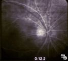
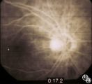
Optic Neuropathies | Larry P. Frohman, MD | This healthy 29-year-old man with dense amblyopia OS presented with a foreign-body sensation OS and further visual loss in his amblyopic eye. He was noted to have bilateral disc edema and lesions in the left eye consistent with unilateral acute multifocal placoid pigment epitheliopathy (AMPPE). He r... |
| 120 |
 |
Optic Neuropathies | Larry P. Frohman, MD | This healthy 29-year-old man with dense amblyopia OS presented with a foreign-body sensation OS and further visual loss in his amblyopic eye. He was noted to have bilateral disc edema and lesions in the left eye consistent with unilateral acute multifocal placoid pigment epitheliopathy (AMPPE). He r... |
| 121 |
 |
Optic Neuropathies | Larry P. Frohman, MD | This healthy 29-year-old man with dense amblyopia OS presented with a foreign-body sensation OS and further visual loss in his amblyopic eye. He was noted to have bilateral disc edema and lesions in the left eye consistent with unilateral acute multifocal placoid pigment epitheliopathy (AMPPE). He r... |
| 122 |
 |
Optic Neuropathies | Larry P. Frohman, MD | This healthy 29-year-old man with dense amblyopia OS presented with a foreign-body sensation OS and further visual loss in his amblyopic eye. He was noted to have bilateral disc edema and lesions in the left eye consistent with unilateral acute multifocal placoid pigment epitheliopathy (AMPPE). He r... |
| 123 |
 |
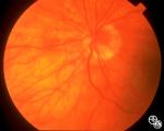
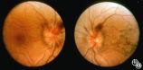
Systemic Disorders With Optic Nerve and Retinal Findings | Larry P. Frohman, MD | At age 41, in 1984, this woman, who grew up in the Ohio River Valley, had 3 days of ocular pain OD, and her vision declined to 20/80 OD she has had no visual changes since, nor has she had any other neurologic symptoms. The ""presumed"" diagnosis is optic neuropathy in presumed ocular histoplasmosis... |
| 124 |
 |
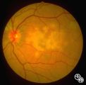
Systemic Disorders With Optic Nerve and Retinal Findings | Larry P. Frohman, MD | This 57-year-old man had a neuro-ophthalmology consult, requested the night before his 2-cm pituitary tumor was to be resected. His examination revealed his acuities to be 20/70 OU, with a visual field not consistent with chiasmal compression. The fundus appearance, with peripheral salt and pepperin... |
| 125 |
 |
Neuro-Ophthalmic Vascular Disease | Larry P. Frohman, MD | This 27-year-old woman had no past ocular history and presented with 3 weeks of redness OS that has been treated by the referring doctor as allergic conjunctivitis. She was referred for evaluation when she developed binocular diplopia. Her past medical history included phlebitis and one miscarriage ... |