Best known for his world-renowned neuro-ophthalmology unit based at the University of California, San Francisco, William Hoyt, MD collected here more than 850 of his best images covering a wide range of disorders.
William F. Hoyt, MD, Professor Emeritus of Ophthalmology, Neurology and Neurosurgery, Department of Ophthalmology, University of California, San Francisco.
NOVEL: https://novel.utah.edu/
TO
Filters: Collection: ehsl_novel_wfh
| Identifier | Title | Description | Subject | ||
|---|---|---|---|---|---|
| 76 |
 |
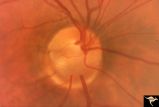
C3_03 | C303 Nodular Papillopathies (Sarcoid) | Lumpy infiltrative papillopathy in a patient with proven sarcoid. Anatomy: Optic disc. Pathology: Axoplasmic stasis due to sarcoid infiltration. Disease/ Diagnosis: Sarcoid papillopathy. Clinical: Unknown? | Disc Swelling; Inflammatory Papillopathies; Nodular Papillopathies; Sarcoid |
| 77 |
 |
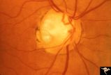
C3_04 | C304 Nodular Papillopathies (Sarcoid) | Lumpy nodular disc infiltration from sarcoid. Anatomy: Optic disc. Pathology: Axoplasmic stasis due to sarcoid infiltration. Disease/ Diagnosis: Sarcoid papillopathy. Clinical: Unknown? | Disc Swelling; Inflammatory Papillopathies; Nodular Papillopathies; Sarcoid |
| 78 |
 |
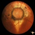
C3_05 | C305 Nodular Papillopathies (Sarcoid) | July 1984 shows multiple infiltrative nodules on the optic disc in addition to circumferential subretinal yellow exudates. 32 year old black woman. Same patient as C3_06 and C3_07. Anatomy: Optic disc; Retina. Pathology: Axoplasmic stasis due to sarcoid infiltration and retinal exudation. Disease/ ... | Disc Swelling; Inflammatory Papillopathies; Nodular Papillopathies; Sarcoid; Sarcoidosis |
| 79 |
 |
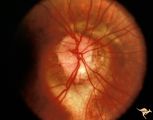
C3_06 | C306 Nodular Papillopathies (Sarcoid) | Lumpy disc swelling with retinal folds and a macular star in a patient with sarcoid. Presentation in October 1983. Same patient as C3_05 and C3_07. Anatomy: Optic disc; Retina. Pathology: Axoplasmic stasis due to sarcoid infiltration. Disease/ Diagnosis: Axoplasmic stasis due to sarcoid infiltration... | Disc Swelling; Inflammatory Papillopathies; Nodular Papillopathies; Sarcoid; Sarcoidosis |
| 80 |
 |
C3_07 | C307 Nodular Papillopathies (Sarcoid) | Fluorescein angiogram shows striking staining of the sarcoid nodules. July 1984. Same patient as C3_05 and C3_06. Corresponds with July 1984 image, C3_06. Anatomy: Optic disc. Pathology: Axoplasmic stasis due to sarcoid infiltration. Disease/ Diagnosis: Sarcoid papillopathy. Clinical: Unknown? | Disc Swelling; Inflammatory Papillopathies; Nodular Papillopathies; Sarcoid; Sarcoidosis |
| 81 |
 |
C3_08 | C308 Nodular Papillopathies (Sarcoid) | Nodular infiltrative papillopathy in a patient with sarcoid. Woman. Anatomy: Optic disc. Pathology: Axoplasmic stasis due to sarcoid infiltration. Disease/ Diagnosis: Sarcoid papillopahty. Clinical: Unknown? | Disc Swelling; Inflammatory Papillopathies - Nodular Papillopathies (Sarcoid) |
| 82 |
 |
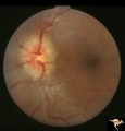
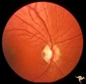
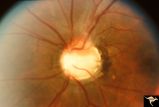
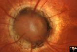
C4_01 | C401 Luetic Papillopathy (Gumma of the Optic Disc) | Diffuse optic disc swelling with tortuous capillary dilations indicating inflammatory cellular infiltration. October 2001. Same eye as C4_02. Anatomy: Optic disc. Pathology: Axoplasmic stasis due to syphillitic infection. Luetic papillopathy (Syphyllis). Clinical: Visual loss. | Disc Swelling; Inflammatory Papillopathies; Luetic Papillopathy; Gumma of the Optic Disc); Syphilis |
| 83 |
 |
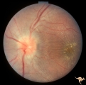
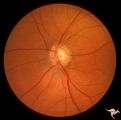
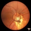
C4_02 | C402 Luetic Papillopathy (Gumma of the Optic Disc) | November 2001. Same eye as C4_01 after treatment with penicillin. Disc swelling went away and good visual function returned. Anatomy: Optic disc. Pathology: Axoplasmic stasis due to syphillitic infection. Disease/ Diagnosis: Luetic papillopathy (Syphillis). Clinical: Improving visual loss. | Disc Swelling; Inflammatory Papillopathies; Luetic Papillopathy (Gumma of the Optic Disc); Syphilis |
| 84 |
 |
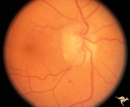
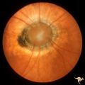
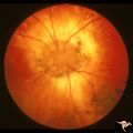
C4_03 | C403 Luetic Papillopathy (Gumma of the Optic Disc) | 40 year old man with AIDS and neurosyphillis with severe visual field defect. The disc is pale and swollen and its arteries are strikingly narrowed (syphillitic vasculitis). Anatomy: Optic disc. Pathology: Axoplasmic stasis due to syphillitic infection. Disease/ Diagnosis: Luetic papillopathy (Syphy... | Disc Swelling; Inflammatory Papillopathies; Luetic Papillopathy; Gumma of the Optic Disc |
| 85 |
 |
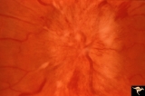
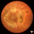
C_01 | C01 Pits of the Optic Disc | Right eye. Very large inferior temporal optic pit. Congenital. Woman. Anatomy: Optic disc. | Cavitary Anomalies; Pits of the Optic Disc |
| 86 |
 |
C_02 | C02 Pits of the Optic Disc | Right eye. Three congenital optic pits on the temporal side. 8:00, 9:30, 10:30. Anatomy: Optic disc. | Cavitary Anomalies; Pits of the Optic Disc |
| 87 |
 |
C_03 | C03 Pits of the Optic Disc | Central optic pit. Left eye. Anatomy: Optic disc. | Cavitary Anomalies; Pits of the Optic Disc |
| 88 |
 |
C_04 | C04 Pits of the Optic Disc | Right eye. Man. Large temporal pit. Macular detachment. Anatomy: Optic disc. | Cavitary Anomalies; Pits of the Optic Disc |
| 89 |
 |
C_05 | C05 Pits of the Optic Disc | Right eye. Pigmented pit. Woman. Anatomy: Optic disc. | Cavitary Anomalies; Pits of the Optic Disc |
| 90 |
 |
C_06 | C06 Pits of the Optic Disc | Right eye. Temporal pit. 6 year old with see-saw nystagmus. Anatomy: Optic disc. Clinical: Six-year old with see-saw nystagmus. | Cavitary Anomalies; Pits of the Optic Disc |
| 91 |
 |
C_07 | C07 Pits of the Optic Disc | Left eye. Temporal pit. Man. Anatomy: Optic disc. | Cavitary Anomalies; Pits of the Optic Disc |
| 92 |
 |
C_08 | C08 Pits of the Optic Disc | Left eye. Large cavitary anomaly (pit). Man. 20/100 visual acuity. Superior nasal visual field defect. May not have a central retinal artery. Anatomy: Optic disc. Clinical: Man. 20/100 visual acuity. Superior nasal visual field defect. | Cavitary Anomalies; Pits of the Optic Disc |
| 93 |
 |
C_09 | C09 Pits of the Optic Disc | Pit with peripapillary choroidal defect. Right eye. Dwarfed boy. May not have a central retinal artery. Same patient as C_10. Anatomy: Optic disc. | Cavitary Anomalies; Pits of the Optic Disc |
| 94 |
 |
C_10 | C10 Pits of the Optic Disc | Disc malformation. Abortive cavitary anomaly. Left eye. Dwarfed boy. Same patient as C_9. Anatomy: Optic disc. | Cavitary Anomalies; Pits of the Optic Disc |
| 95 |
 |
C_11 | C11 Morning Glory Disc | "Morning Glory" disc. 11 year old girl. May not have a central retinal artery. Anatomy: Optic disc. | Cavitary Anomalies; Morning Glory Disc; Morning Glory Disc Anomaly; Morning Glory Syndrome |
| 96 |
 |
C_12 | C12 Morning Glory Disc | "Morning Glory" disc. Patient 11 year old. Anatomy: Optic disc. | Cavitary Anomalies; Morning Glory Disc |
| 97 |
 |
C_13 | C13 Morning Glory Disc | "Morning Glory" disc with peripapillary choroidal defect extending inferiorly. Patient has transphenoidal encephalocele. Note tapering edge like an arrow pointing to patient's basal encephalocele and cleft palate. Reference: Brodsky MC, Hoyt WF, Hoyt CS, Miller NR, Lam BL. Atypical retinochoroidal ... | Cavitary Anomalies; Morning Glory Disc |
| 98 |
 |
C_14 | C14 Morning Glory Disc | Isolated "Morning Glory". Left eye. Girl. Anatomy: Optic disc. | Cavitary Anomalies; Morning Glory Disc |
| 99 |
 |
C_15 | C15 Morning Glory Disc | "Morning Glory" disc. Note tapering edge pointing to patient's transphenoidal encephalocele. Reference: Brodsky MC, Hoyt WF, Hoyt CS, Miller NR, Lam BL. Atypical retinochoroidal coloboma in patients with dysplastic optic discs and transphenoidal encephalocele Arch Ophthalmol. 1995 May;113(5):624-8.... | Cavitary Anomalies; Morning Glory Disc |
| 100 |
 |
C_16 | C16 Morning Glory Disc | "Morning Glory" disc. Note tapering edge pointing to basal encephalocele. Boy. Anatomy: Optic disc. | Cavitary Anomalies; Morning Glory Disc |