A collection of videos relating to the diagnosis and treatment of eye movement disorders. This collection includes many demonstrations of examination techniques.
Dan Gold, D.O., Associate Professor of Neurology, Ophthalmology, Neurosurgery, Otolaryngology - Head & Neck Surgery, Emergency Medicine, and Medicine, The Johns Hopkins School of Medicine.
A collection of videos relating to the diagnosis and treatment of eye movement disorders.
NOVEL: https://novel.utah.edu/
TO
Filters: Collection: ehsl_novel_gold
| Title | Description | Subject | ||
|---|---|---|---|---|
| 76 |
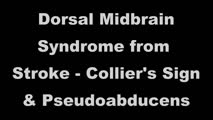 |
Dorsal Midbrain Syndrome from Stroke - Collier's Sign & Pseudoabducens | This is a 70-yo-man who suffered a right midline thalamic/rostral midbrain hemorrhagic stroke causing a pretectal (Parinaud's) syndrome. There was prominent eyelid retraction (Collier's sign), a left pseudo-abducens, and upgaze palsy with convergence retraction nystagmus. There was no light-near dis... | OMS Mesenecephalon; Convergence Reaction Nystagmus; Upgaze Palsy; Abnormal Range; Sixth Nerve Palsy; Vertical Gaze Palsy; Eyelid Retraction; OMS Dorsal Midbrain |
| 77 |
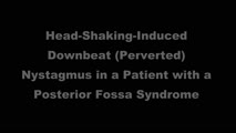 |
Downbeat (Perverted) Head Shaking Nystagmus in a Patient with Spontaneous Torsional Nystagmus | 𝗢𝗿𝗶𝗴𝗶𝗻𝗮𝗹 𝗗𝗲𝘀𝗰𝗿𝗶𝗽𝘁𝗶𝗼𝗻: This is a 75-year-old woman with vascular risk factors who experienced abrupt onset imbalance and dizziness. Symptoms were maximal at onset, and she denied progression over 6 months. Clinically, it was felt that she had s... | Torsional Nystagmus; Jerk Nystagmus; Headshaking; Downbeat Nystagmus; Cerebellar Pathology |
| 78 |
 |
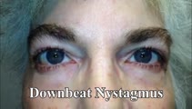
Downbeat Nystagmus | 𝗢𝗿𝗶𝗴𝗶𝗻𝗮𝗹 𝗗𝗲𝘀𝗰𝗿𝗶𝗽𝘁𝗶𝗼𝗻: This is a 40-year-old man with 2 years of progressive ataxia and oscillopsia. On examination, he had downbeat nystagmus (DBN), an ocular motor finding that is usually (but not always) associated with flocculus/parafloccul... | Downbeat Nystagmus; OMS Cerebellar; Jerk Nystagmus; Vestibular Nystagmus |
| 79 |
|
Downbeat Nystagmus and Cerebellar Atrophy | This is a 40-year-old man with 2 years of progressive ataxia and oscillopsia. On examination, he had downbeat nystagmus (DBN), an ocular motor finding that is usually (but not always) associated with flocculus/paraflocculus dysfunction, which causes overaction of the anterior canal (upward or anti-g... | Downbeat Nystagmus; OMS Cerebellar; Jerk Nystagmus; Vestibular Nystagmus |
| 80 |
 |
Downbeat Nystagmus and Convergence Spasm | This is a 60-yo-woman with vertical oscillopsia related to her downbeat nystagmus, and diplopia related to an intermittent esotropia. When the esotropia was present, with versions there were bilateral abduction deficits. With ductions and the vestibulo-ocular reflex, it was apparent that the range o... | Convergence-abnormal; Range-abnormal; Sixth Nerve Palsy; Convergence Spasm; Cerebellar Pathology; Jerk Nystagmus; Downbeat Nystagmus |
| 81 |
 |
Downbeat Nystagmus with Active Horizontal Head Shaking | This is a 70-year-old man who presented with one single complaint for 10 years - if he moved his head too quickly (even one single horizontal head movement to the right or the left), he would experience the abrupt loss of balance and dizziness. His typical episodes were reproducible, and interesting... | Downbeat Nystagmus; Jerk Nystagmus; OMS Cerebellar; Abnormal Headshaking; Gaze-Evoked Nystagmus |
| 82 |
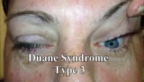 |
Duane's Syndrome Type III | This is a 40-yo-woman seen in neurology clinic for a complaint unrelated to her eyes. On exam, there was impaired adduction and abduction OS. In adduction, there was narrowing of the palpebral fissure OS, a result of her globe retraction due to co-contraction of the medial and lateral rectus muscles... | Duane's Syndrome; Type 3 |
| 83 |
 |
Dynamic Visual Acuity | 𝗢𝗿𝗶𝗴𝗶𝗻𝗮𝗹 𝗗𝗲𝘀𝗰𝗿𝗶𝗽𝘁𝗶𝗼𝗻: After assessing static binocular visual acuity, dynamic visual acuity (DVA) is determined by repeating the test during horizontal and vertical head shaking at 2-3 Hz. Dynamic visual acuity is most important to test when ... | Dynamic Visual Acuity |
| 84 |
 |
Dynamic Visual Acuity | Dynamic Visual Acuity: the examiner can use screen-sharing to provide a visual acuity chart. Instruct the patient to sit at the appropriate distance from their screen at which the lowest line on the visual acuity chart is just readable. Have the patient move their head (horizontally to evaluate the ... | Dynamic Visual Acuity |
| 85 |
 |
ENG, VNG, & VOG | 𝗢𝗿𝗶𝗴𝗶𝗻𝗮𝗹 𝗗𝗲𝘀𝗰𝗿𝗶𝗽𝘁𝗶𝗼𝗻: Electronystagmography (ENG), and videonystagmography (VNG) or videooculography (VOG) are a collection of tests of eye movements that are performed either using surface electrodes around the eye (ENG) or with video goggles... | Vestibular Lab Testing; ENG, VNG, & VOG |
| 86 |
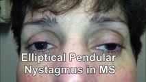 |
Elliptical Pendular Nystagmus in MS | 𝗢𝗿𝗶𝗴𝗶𝗻𝗮𝗹 𝗗𝗲𝘀𝗰𝗿𝗶𝗽𝘁𝗶𝗼𝗻: This is a 40-yo-woman with MS and bilateral optic nerve disease who presented with a year's long history of oscillopsia, which was related to elliptical pendular nystagmus. The appearance of elliptical nystagmus is the re... | Pendular Nystagmus; Multiple Sclerosis |
| 87 |
 |
Enhanced Ptosis in Myasthenia Gravis | This is a 20-yo-woman who presented with generalized weakness, ptosis and ophthalmoplegia. She had severe ptosis OU at baseline, but when one eyelid was manually elevated, there was marked enhanced ptosis of the opposite eyelid. This was in accordance with Hering's law of equal innervation of the le... | Myasthenia Gravis |
| 88 |
 |
Evaluation of Auditory Function Using Rinne and Weber Tests | 𝗢𝗿𝗶𝗴𝗶𝗻𝗮𝗹 𝗗𝗲𝘀𝗰𝗿𝗶𝗽𝘁𝗶𝗼𝗻: The Rinne test is an assessment of auditory thresholds to air and bone conduction of sound. The Weber test is a comparison of bone conducted sound of either ear. Conductive hearing loss results in a loss of air conducte... | Auditory Testing |
| 89 |
 |
Evaluation of Convergence | 𝗢𝗿𝗶𝗴𝗶𝗻𝗮𝗹 𝗗𝗲𝘀𝗰𝗿𝗶𝗽𝘁𝗶𝗼𝗻: The assessment of convergence includes measuring alignment at near versus distance (see video, https://collections.lib.utah.edu/details?id=187677), near point of convergence and convergence amplitude. Near point of conve... | Normal Convergence |
| 90 |
 |
Examples of Patients with Saccadic Intrusions (Square Wave Jerks) | Seen here are patients with saccadic intrusions that do have an intersaccadic interval. Square wave jerks are commonly seen in degenerative conditions, mainly involving the posterior fossa (e.g., cerebellar degeneration) and basal ganglia (e.g., progressive supranuclear palsy). | Square Wave Jerks |
| 91 |
 |
Expanded Acute Onset Persistent Vision Loss Differential | ||
| 92 |
 |
Expanded Nystagmus & Saccadic Intrusions/Oscillations Differential | Expanded nystagmus & saccadic intrusions/ oscillations differential | |
| 93 |
 |
Eye Closure and Oculopalatal Tremor | 𝗢𝗿𝗶𝗴𝗶𝗻𝗮𝗹 𝗗𝗲𝘀𝗰𝗿𝗶𝗽𝘁𝗶𝗼𝗻: This patient suffered a traumatic brain injury with brainstem injury resulting in damage to Mollaret's triangle and palatal tremor. Inferior olivary hypertrophy was noted on her MRI, although no vertical and/or torsional ... | Pendular Nystagmus; Oculopalatal Tremor |
| 94 |
 |
Eye Handbook App for OKN | Optokinetic nystagmus (OKN): one way this can be examined virtually is using a smartphone application (e.g. Eye Handbook © app used in this video) or optokinetic tape/flag/drum held in front of the examiner's camera. The optokinetic stimulus should occupy the full screen of the patient's device (ea... | Eye Handbook App for OKN |
| 95 |
 |
Eye Signs in Infantile Esotropia - Latent Nystagmus and Inferior Oblique Overaction | 𝗢𝗿𝗶𝗴𝗶𝗻𝗮𝗹 𝗗𝗲𝘀𝗰𝗿𝗶𝗽𝘁𝗶𝗼𝗻: This is a 25-yo-man with a history of amblyopia and intermittent eye crossing. On exam, he had a comitant 25 prism diopter esotropia, and other features of infantile (or congenital) esotropia including: latent nystagmus (... | Jerk Nystagmus; Latent Nystagmus; Abnormal Range; Assessing Abnormal Alignment |
| 96 |
 |
Eyelid Anatomy | Seen here are the major muscles of eyelid opening and closure. The levator palpebrae, which is innervated by the oculomotor nerve, inserts on the tarsus via the levator aponeurosis and directly on the skin of the upper eyelid. The superior tarsal muscle (also known as Muller's muscle, which is inner... | Figures |
| 97 |
 |
Eyelid Nystagmus | Lid nystagmus is a rhythmic eyelid movement commonly seen as an epiphenomenon of vertical nystagmus (typically upbeating, as in this case) due to a shared central pathway controlling elevation of the lid and supraduction. There can be isolated lid nystagmus if there is accompanying impairment of su... | Nystagmus; Eyelid Nystagmus |
| 98 |
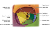 |
Figure 17: Bony Structures Relevant to the Orbit | The frontal, sphenoid, maxillary, ethmoid, and lacrimal bones make up the orbit. Structures passing through the optic canal include the optic nerve, oculosympathetic tract and ophthalmic artery. Structures passing through the superior orbital fissure include the superior ophthalmic vein, cranial ner... | |
| 99 |
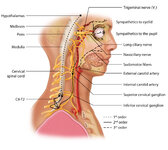 |
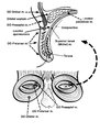
Figure 1: Oculosympathetic Pathway for Pupillary Dilation | The oculosympathetic tract is an uncrossed pathway that begins in the hypothalamus, with fibers descending in the brainstem (1st order, commonly affected in a lateral medullary syndrome), synapsing in the lower cervical/upper thoracic spinal cord (interomediolateral cell columns of C8-T2, also refer... | |
| 100 |
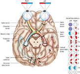 |
Figure 24: Typical Visual Field Defects Associated with Discrete Lesions Along the Visual Pathways | Specific monocular or binocular visual field defects can be highly localizing when the neuroanatomy of the visual pathways is understood. The temporal visual field corresponds to the nasal retina, while the nasal visual field corresponds to the temporal retina. 1) Left optic nerve lesion - while an ... |