Best known for his world-renowned neuro-ophthalmology unit based at the University of California, San Francisco, William Hoyt, MD collected here more than 850 of his best images covering a wide range of disorders.
William F. Hoyt, MD, Professor Emeritus of Ophthalmology, Neurology and Neurosurgery, Department of Ophthalmology, University of California, San Francisco.
NOVEL: https://novel.utah.edu/
TO
| Title | Description | Type | ||
|---|---|---|---|---|
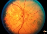
| 76 |
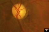 |
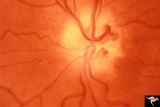
Diffuse Atrophy | Primary optic atrophy from optic nerve compression by aneurysm. Note narrowing of retinal arterioles. Pair with IIA1_8. Anatomy: Optic disc. Pathology: Optic atrophy. Disease/Diagnosis: Optic atrophy due to giant aneurysm. Clinical: Blindness. | Image |
| 77 |
 |
Diffuse Atrophy - Evolution of Optic Disc Palor After Optic Nerve Transection | Evolution of optic disc pallor after optic nerve transection. Normal Right eye. Photo taken December 9, 1978. Anatomy: Optic disc. Pathology: Total retrograde optic atrophy. Disease/Diagnosis: Transection of the optic nerve. Clinical: Blindness. | Image |
| 78 |
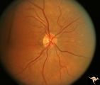
 |
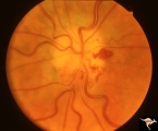
Diffuse Atrophy - Evolution of Optic Disc Palor After Optic Nerve Transection | Injury on December 8, 1978. Evolution of optic disc pallor after optic nerve transection. Woman having rhinoplasty suffered optic nerve transection. Left eye. Photo taken January 11, 1979 - 33 days post accident. Note superior and inferior arcuate nerve fiber bundles are thinned. Optic disc shows s... | Image |
| 79 |
 |
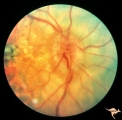
Diffuse Atrophy - Evolution of Optic Disc Palor After Optic Nerve Transection | Injury on December 8, 1978. Evolution of optic disc pallor after optic nerve transection. Woman having rhinoplasty suffered optic nerve transection. Left eye. Photo taken February 14, 1979 - 65 days post accident. Optic disc is completely pale. All evidence of retinal nerve fiber layer is gone. Anat... | Image |
| 80 |
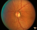
 |
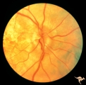
Diffuse Atrophy - Evolution of Optic Disc Palor After Optic Nerve Transection | Injury on December 8, 1978. Evolution of optic disc pallor after optic nerve transection. Woman having rhinoplasty suffered optic nerve transection. Left eye. Photo taken January 18, 1979 - 40 days post accident. Retinal nerve fiber layer appears thinner and disc is paler. Anatomy: Optic disc. Patho... | Image |
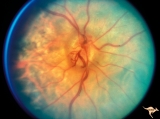
| 81 |
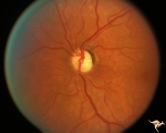
 |
Diffuse Atrophy - Evolution of Optic Disc Palor After Optic Nerve Transection | Injury on December 8, 1978. Evolution of optic disc pallor after optic nerve transection. Woman having rhinoplasty suffered optic nerve transection. One day after nerve transection. Note dilated veins. Left eye. Photo taken December 9, 1978. Anatomy: Optic disc. Pathology: Total retrograde optic atr... | Image |
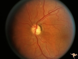
| 82 |
 |
F207 Disc Swelling due to Metastatic Breast Cancer | Unilateral disc swelling with retinal folds due to metastatic breast cancer. Apparent enophthalmus. Anatomy: Optic disc. Pathology: Metastatic breast cancer. Disease/ Diagnosis: Neoplastic papillopathy. | Image |
| 83 |
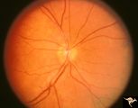 |
H53 Superior Segmental Optic Hypoplasia (SSOH) Topless Disc Syndrome | Superior segmental optic hypoplasia. High exit point of central retinal vessels. Anatomy: Optic disc. Pathology: Superior segmental optic hypoplasia (SSOH). Disease/ Diagnosis: Superior segmental optic hypoplasia (SSOH). | Image |
| 84 |
 |
H54 Superior Segmental Optic Hypoplasia (SSOH) Topless Disc Syndrome | Note superior segmental palor. Anatomy: Optic disc. Pathology: Superior segmental optic hypoplasia (SSOH). Disease/ Diagnosis: Congenital anomaly. | Image |
| 85 |
 |
H55 Superior Segmental Optic Hypoplasia (SSOH) Topless Disc Syndrome | Note superior choroidal crescent. Anatomy: Optic disc. Pathology: Superior segmental optic hypoplasia (SSOH). Disease/ Diagnosis: Congenital anomaly. | Image |
| 86 |
 |
H56 Superior Segmental Optic Hypoplasia (SSOH) Topless Disc Syndrome | High exit point of central retinal vessels. Anatomy: Optic disc. Pathology: Superior segmental optic hypoplasia (SSOH). DIsease/ Diagnosis: Congenital anomaly. | Image |
| 87 |
 |
H89 Occipital Hemianoptic Hypoplasia | Diagram of homonymous hemioptic hypoplasia showing pattern of preserved nerve fibers. Homonymous hemioptic hypoplasia. Fundoscopic features in standard and red-free illumination in three patients with congenital hemiplegia. Anatomy: Optic disc. Pathology: Occipital hemianoptic hypoplasia. Disease/ D... | Image |
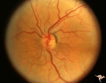
| 88 |
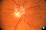 |
IA01 Atrophy with Optociliary Veins | 1994, perioptic nerve sheath meningioma, right eye, Optociliary vein dumping into disc edge at 4:00. Anatomy: Optic disc. Pathology: Optociliary vein. Disease/ Diagnosis: Perioptic nerve sheath meningioma. Clinical: Progressive visual loss | Image |
| 89 |
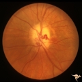 |
IA02 Atrophy with Optociliary Veins | 1971, left eye, perioptic nerve sheath meningioma, notice how vein dumps into adjacent choroid at 3:00. The darker venous blood can be seen at the disc edge. Anatomy: Optic disc. Pathology: Optociliary vein. Disease/ Diagnosis: Perioptic nerve sheath meningioma. Clinical: Progressive visual loss. | Image |
| 90 |
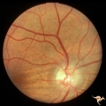 |
IA04 Atrophy with Optociliary Veins | 1981, right eye, perioptic nerve sheath meningioma with optociliary bypass vein. Notice horizontal choroidal folds in the retina from posterior tumor pressure. Anatomy: Optic disc. Pathology: Optociliary vein. Disease/ Diagnosis: Perioptic nerve sheath meningioma. Clinical: Blind eye. | Image |
| 91 |
 |
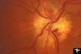
IA05 Atrophy with Optociliary Veins | 1971, right eye, perioptic nerve sheath meningioma with optociliary bypass veins on the upper half of the disc. Anatomy: Optic disc. Pathology: Optociliary vein. Disease/ Diagnosis: Perioptic nerve sheath meningioma. Clinical: Blind eye. | Image |
| 92 |
 |
IA06 Atrophy with Optociliary Veins | 1979, left eye, perioptic nerve sheath meningioma with optociliary bypass veins. Anatomy: Optic disc. Pathology: Optociliary vein. Disease/ Diagnosis: Perioptic nerve sheath meningioma. Clinical: Blind eye. | Image |
| 93 |
 |
IA07 Atrophy with Optociliary Veins | Left eye, perioptic nerve sheath meningioma. Anatomy: Optic disc. Pathology: Optociliary vein. Disease/ Diagnosis: Perioptic nerve sheath meningioma. Clinical: Visual loss. | Image |
| 94 |
 |
IA08 Atrophy with Optociliary Veins | 1996, left eye. Chronic pale optic nerve swelling with optociliary bypass veins produced by perioptic nerve sheath meningioma. Anatomy: Optic disc. Pathology: Optociliary vein. Disease/ Diagnosis: Perioptic nerve sheath meningioma evolution. Clinical: Visual loss. | Image |
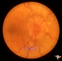
| 95 |
 |
IA09a Evolution of Optociliary Veins with Perioptic Nerve Sheath Meningioma | April 1975, Normal eye, macular degeneration. Anatomy: Optic disc. Pathology: Optociliary vein. Disease/ Diagnosis: Perioptic nerve sheath meningioma evolution. Clinical: Visual loss. | Image |
| 96 |
 |
IA09b Evolution of Optociliary Veins with Perioptic Nerve Sheath Meningioma | January 1977, macular degeneration, disc swelling begins. Anatomy: Optic disc. Pathology: Optociliary vein. Disease/ Diagnosis: Perioptic nerve sheath meningioma evolution. Clinical: Visual loss. | Image |
| 97 |
 |
IA09c Evolution of Optociliary Veins with Perioptic Nerve Sheath Meningioma | June 1977, continued disc swelling. Anatomy: Optic disc. Pathology: Optociliary vein. Disease/ Diagnosis: Perioptic nerve sheath meningioma evolution. Clinical: Visual loss. | Image |
| 98 |
 |
IA09d Evolution of Optociliary Veins with Perioptic Nerve Sheath Meningioma | October 1977, continued disc swelling. Anatomy: Optic disc. Pathology: Optociliary vein. Disease/ Diagnosis: Perioptic nerve sheath meningioma evolution. Clinical: Visual loss. | Image |
| 99 |
 |
IA09e Evolution of Optociliary Veins with Perioptic Nerve Sheath Meningioma | February 1979, development of optociliary veins at 7:00, 1:00. Anatomy: Optic disc. Pathology: Optociliary vein. Disease/ Diagnosis: Perioptic nerve sheath meningioma evolution. Clinical: Visual loss. | Image |
| 100 |
 |
IA09f Evolution of Optociliary Veins with Perioptic Nerve Sheath Meningioma | August 1979, less disc swelling and development of atrophy with more prominent optociliary veins at 7:00 and 1:00. Anatomy: Optic disc. Pathology: Optociliary vein. Disease/ Diagnosis: Perioptic nerve sheath meningioma evolution. Clinical: Visual loss. | Image |