Best known for his world-renowned neuro-ophthalmology unit based at the University of California, San Francisco, William Hoyt, MD collected here more than 850 of his best images covering a wide range of disorders.
William F. Hoyt, MD, Professor Emeritus of Ophthalmology, Neurology and Neurosurgery, Department of Ophthalmology, University of California, San Francisco.
NOVEL: https://novel.utah.edu/
TO
| Title | Description | Type | ||
|---|---|---|---|---|
| 76 |
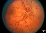 |
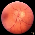
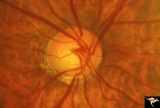
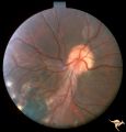
E04 Disc Swelling with Central Retinal Vein Occlusion | Acute CRVO, right eye with disc swelling. Male patient. Same patient as E05. Anatomy: Optic disc; Retina. Pathology: Central retinal vein occlusion. Disease/ Diagnosis: Disc swelling due to central retinal vein occlusion. Clinical: Visual blurring. | Image |
| 77 |
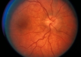 |
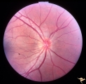
E05 Disc Swelling with Central Retinal Vein Occlusion | Resolving CVRO, right eye. Two months following slide E04. Male patient. Anatomy: Optic disc; Retina. Pathology: Central retinal vein occlusion. Disease/ Diagnosis: Resolved disc swelling after central retinal vein occlusion. Clinical: No symptoms. | Image |
| 78 |
 |
Crowded Disc (Family) | Anomalous vasculature with congenital disc margin blurring. Note optic cup is absent. Pair with brother in PP1a & b. Mother has drusen of the optic disc in PP11aa & b. Sister has drusen in PP11c. Anatomy: Optic disc. Pathology: Normal variant. Cause of appearance is too many fibers entering into a s... | Image |
| 79 |
 |
PP8a Crowded Disc with Significant Nasal Disc Blurring | Congenital nasal disc blurring. Myopic eyes. Thai girl patient. One wonders about vitreal adherence to the disc. PP 8a right eye. Pair with left eye in PP8b. Anatomy: Optic disc. Pathology: Normal variation of the optic disc. Disease/ Diagnosis: Normal variation of the optic disc. Congenital blurre... | Image |
| 80 |
 |
Unilateral Pseudopapilledema | PP_10a: Left: pseudo papilledema with disc blurring, crowded disc. Optic disc is small in diameter. PP_10b shows albinotic fundus and small crowded disc. Anatomy: Optic disc. Pathology: Normal variation of the optic disc. Disease/Diagnosis: Normal variation of the optic disc. Elevated disc. Clinic... | Image |
| 81 |
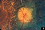 |
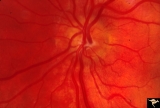
Visible Drusen with Retinitis Pigmentosa | Right eye. Optic disc drusen with retinitis pigmentosa. Note the marked narrowing of the retinal arterioles and the spectacular change in the peripapillary choroid. Anatomy: Optic disc. Pathology: Drusen of the optic disc. Disease/Diagnosis: Drusen of the optic disc. Clinical: Patient was nearly bli... | Image |
| 82 |
 |
Drusen Plus Papilledema | PP37a: right swollen disc on top of drusen with narrowing of the arterioles; PP37b: left visible drusen and papilledema with sub-retinal hemorrhage temporally. Patient had frontal glioblastoma. Anatomy: Optic disc. Pathology: Drusen of the optic disc. Disease/Diagnosis: Drusen of the optic disc. Cli... | Image |
| 83 |
 |
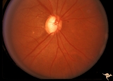
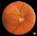
Congenitally Crowded Disc - Little Red Disc | Right eye: "little red disc". Congenitally blurred disc. 26 year old man. Anatomy: Optic disc Pathology: Normal variation of the optic disc Disease/Diagnosis: Normal variation of the optic disc. Congenital blurred disc. Little red disc. | Image |
| 84 |
 |
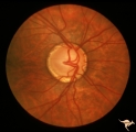
IF107 Glaucoma Cupped Disc | Glaucoma cupped disc with inferior temporal retinal nerve fiber layer defect. Vertically ovoid cup. 1974. Anatomy: Optic disc. Pathology: Glaucoma. Disease/ Diagnosis: Glaucoma. Clinical: Superior arcuate visual field defects. | Image |
| 85 |
 |
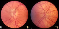
IF102a Low Tension Glaucoma | Low tension glaucoma with bilateral superior altitudinal field defects. Thinning of the neuroretinal rim. Cupping predominantly inferiorly. Pair with IF1_2b. Anatomy: Optic disc. Pathologhy: Glaucoma. Disease/ Diagnosis: Low tension glaucoma. Clinical: Bilateral altitudinal visual field loss. | Image |
| 86 |
 |
IF102b Low Tension Glaucoma | Low tension glaucoma with bilateral superior altitudinal field defects. Thinning of the neuroretinal rim. Cupping predominantly inferiorly. Pair with IF1_2b. Anatomy: Optic disc. Pathology: Glaucoma. Disease/ Diagnosis: Low tension glaucoma. Clinical: Bilateral altitudinal visual field loss. | Image |
| 87 |
 |
IF104b Low Tension Glaucoma | Possible low tension glaucoma. Patient with macro discs with remarkable cupping. Pair with IF1_4a. 1969. Anatomy: Optic disc. Disease/ Diagnosis: Cupping and megalopapilla (macrodisc). Clinical: Possible visual field defect. | Image |
| 88 |
 |
IF104a Low Tension Glaucoma | Possible low tension glaucoma. Patient with macro discs with remarkable cupping. Pair with IF1_4b. 1969. Anatomy: Optic disc. Disease/ Diagnosis: Cupping and megalopapilla (macrodisc). Clinical: Possible visual field defect. | Image |
| 89 |
 |
IF105a Low Tension Glaucoma | 40 year old man. Megalopapilla. Right eye has superior arcuate field defect. Pair with IF1_5b. Anatomy: Optic disc. Disease/ Diagnosis: Cupping and megalopapilla (macrodisc). Clinical: Asymptomatic. | Image |
| 90 |
 |
IF111d Low Tension Glaucoma | Low tension glaucoma. Followed. Inferior arcuate field defect has expanded upward. Note increase in atrophy and cupping in inferior temporal disc. Pair with IF1_11a, b, d. Right eye. 1990. Anatomy: Optic disc. Pathology: Glaucoma. Disease/ Diagnosis: Low tension glaucoma. Clinical: Increased size o... | Image |
| 91 |
 |
IF111b Low Tension Glaucoma | Low tension glaucoma. Followed, 9 years later. Wedge defects in retinal nerve fiber defects in both temporal arcuate zones. Note small disc edge hemorrhage at 5:00. Pair with IF1_11a, c, d. Left eye. 1990. Anatomy: Optic disc. Pathology: Glaucoma. Disease/ Diagnosis: Low tension glaucoma. Clinical:... | Image |
| 92 |
 |
IF111c Low Tension Glaucoma | Low tension glaucoma. Followed. Notice disc edge hemorrhage at 7:00. Inferior nerve fiber layer defect between 6:00 and 7:30.Pair with IF1_11a, b, d. Right eye. 1981. Anatomy: Optic disc. Pathology: Glaucoma. Disease/ Diagnosis: Low tension glaucoma. Clinical: Superior arcuate visual field defect | Image |
| 93 |
 |
IF105b Low Tension Glaucoma | 40 year old man. Megalopapilla. Left eye. Pair with IF1_5a. Anatomy: Optic disc. Disease/ Diagnosis: Cupping and megalopapilla (macrodisc). Clinical: Asymptomatic. | Image |
| 94 |
 |
IF111a Low Tension Glaucoma | Low tension glaucoma. Followed. Pair with IF1_11b, c, d. Left eye. 1981. Anatomy: Optic disc. Pathology: Glaucoma. Disease/ Diagnosis: Low tension glaucoma. Clinical: Asymptomatic. | Image |
| 95 |
 |
C305 Nodular Papillopathies (Sarcoid) | July 1984 shows multiple infiltrative nodules on the optic disc in addition to circumferential subretinal yellow exudates. 32 year old black woman. Same patient as C3_06 and C3_07. Anatomy: Optic disc; Retina. Pathology: Axoplasmic stasis due to sarcoid infiltration and retinal exudation. Disease/ ... | Image |
| 96 |
 |
C307 Nodular Papillopathies (Sarcoid) | Fluorescein angiogram shows striking staining of the sarcoid nodules. July 1984. Same patient as C3_05 and C3_06. Corresponds with July 1984 image, C3_06. Anatomy: Optic disc. Pathology: Axoplasmic stasis due to sarcoid infiltration. Disease/ Diagnosis: Sarcoid papillopathy. Clinical: Unknown? | Image |
| 97 |
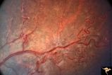 |
Neurofibromatosis-1 | Extensive retinal microvascular malformation involving both small and large retinal vessels. (Ref: BJO 2002:86, p282-284). Anatomy: Retina. Pathology: Retinal microvascular malformations. Disease/Diagnosis: Neurofibromatosis type 1. Clinical: No visual symptoms. | Image |
| 98 |
 |
Neurofibromatosis-2 | CPERH (choroidal pigment epithelial retinal hamartoma) lesion in a patient with NF-2. Note the oblique superficial retinal traction folds running toward the center of the main lesion. 51 year old man. Anatomy: Retina. Pathology: Hamartoma. Disease/Diagnosis: Neurofibromatosis type 2. Clinical: Fiel... | Image |
| 99 |
 |
Neurofibromatosis-2 | Retinal tumor in NF-2 referred to as a CPERH (choroidal pigment epithelial retinal hamartoma). Patient, a 16 year old girl, had bilateral acoustic neurinomas. Pair with R1_F2b. Same eye. Anatomy: Optic disc; Retina. Pathology: Retinal hamartoma; Bilateral acoustic neurinoma. Disease/Diagnosis: Neuro... | Image |
| 100 |
 |
Tuberous Sclerosis | Soft translucent lesion of tuberous sclerosis in the inferior temporal retina. Patient was 4 years old. Anatomy: Retina. Pathology: Astrocytic hamartoma. Disease/Diagnosis: Tuberous sclerosis. Clinical: No visual symptoms. | Image |