Collection of materials relating to neuro-ophthalmology as part of the Neuro-Ophthalmology Virtual Education Library.
NOVEL: https://novel.utah.edu/
TO
- NOVEL720
| Title | Creator | Description | Subject | ||
|---|---|---|---|---|---|
| 76 |
 |
Bergmeister Papilla | Sumayya Almarzouqi, MD | A brief overview of Bergmeister papilla, a rare congenital disc anomaly. It arises from the center of the optic disc consists of a small tuft of fibrous tissue and represents a remnant of the fetal hyaloid artery. | Bergmeister Papilla |
| 77 |
 |
Modern Imaging of Optic Disc Drusen | Meagan Seay, DO | This is a short powerpoint describing imaging techniques (specifically OCT-EDI, fundus autofluorescence, and B-scan ultrasonography) for optic disc drusen. Examples of these techniques are included. | Optic Disc Drusen; Imaging; OCT-EDI; Fundus Autofluorescence; B-scan Ultrasonography |
| 78 |
 |
Suprasellar Meningioma | Sumayya Almarzouqi, MD | Description of a case of suprasellar or sellar mass causeing chiasmal compression. | Suprasellar Meningioma |
| 79 |
 |
Anaesthesia for Eye Surgery and Associated Complications Slides | Julie Smith, MBBS, FANZCA | Lecture covering commonly performed eye surgery and anaesthetic techniques. | Eye Surgery; Anesthesia |
| 80 |
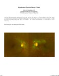 |
Myelinated Retinal Nerve Fibers | Scott N. Grossman, MD | A 33 year old man has noted chronically poor vision OS - left eye color noted to be 'orange' instead of red. fundus photos revealed myelinated retinal nerve fiber layer OU (OS>OD) with corresponding linear paracentral scotoma on Humphrey visual field 24-2 OS corresponding with greatest degree of my... | Myelinated Retinal Nerve Fibers |
| 81 |
 |
Cerebellar Anatomy on MRI | Joshua East, MD; Nicholas A. Koontz, MD; Devin D. Mackay, MD | Overview of structural anatomy of the cerebellum and surround structures on MRI images of the brain. | Cerebellar Anatomy; MRI |
| 82 |
 |
Progressive Supranuclear Palsy (PSP) | Molly Cincotta, MD; Ali G. Hamedani, MD, MHS | Objectives:; To provide an overview of PSP and its pathophysiology;; To present typical clinical features of the disease with a focus on ocular findings;; To provide a template for work up, diagnosis and treatment; ; To demonstrate typical eye movement abnormalities seen in PSP | Progressive Supranuclear Palsy (PSP) |
| 83 |
 |
Myotonic Dystrophy | Brian Villafuerte, MD, Ezequiel Piccione, MD | Presentation covering an overview of myotonic dystrophy. | Myotonic Dystrophy |
| 84 |
 |
Muscular Dystrophy Classification | Brian Villafuerte, MD, Ezequiel Piccione, MD | Presentation covering an overview of muscular dystrophy classification. | Muscular Dystrophy Classification |
| 85 |
 |
Congenital Hydrocephalus | Mays El-Dairi, MD | Presentation covering an overview of congenital hydrocephalus. | Congenital Hydrocephalus |
| 86 |
 |
Fibrous Dysplasia | Mays El-Dairi, MD | Presentation covering an overview of fibrous dysplasia. | Fibrous Dysplasia |
| 87 |
 |
Heavy Eye Syndrome | Meagan D. Seay, DO; Bradley J. Katz, MD | A brief overview of heavy eye syndrome. | Heavy Eye Syndrome |
| 88 |
 |
Brown Syndrome | Meagan Seay, DO | A brief overview of Brown Syndrome. | Brown Syndrome |
| 89 |
 |
Posterior Cortical Atrophy | Natali V. Baner, MD; Ali G. Hamedani, MD, MHS | PowerPoint providing an overview of the definition, clinical presentation and treatment of posterior cortical atrophy | Posterior Cortical Atrophy |
| 90 |
 |
Dementia: Overview and Classification | Molly Cincotta, MD; Whitley Aamodt, MD; Ali G. Hamedani, MD, MHS | PowerPoint providing a broad overview of dementia, including definition, clinical findings, work up, diagnosis, classification, and management. | Dementia |
| 91 |
 |
Multiple System Atrophy: Overview and Neuro-ophthalmologic Features | Pavan Vaswani, MD, PhD, Movement Disorders Fellow; Ali G. Hamedani, MD, MHS | Objectives: Know the key pathologic features of Multiple System Atrophy; Recognize the clinical presentation, including neuro-ophthalmologic features; Understand the symptomatic therapies and prognosis | Multiple System Atrophy |
| 92 |
 |
Dementia with Lewy Bodies: Overview and Neuro-ophthalmologic features | Pavan Vaswani, MD, PhD; Ali G. Hamedani, MD, MHS | Objectives: Recognize the difference between Dementia with Lewy Bodies and Parkinson disease dementia; Recognize the clinical presentation of DLB and differentiating features from Alzheimer disease dementia; Understand the symptomatic therapies and prognosis | Dementia; Lewy Bodies |
| 93 |
 |
Vascular Dementia | Whitley Aamodt, MD; Ali G. Hamedani, MD, MHS | PowerPoint providing an overview of vascular dementia, including the pathophysiology, clinical symptoms, diagnosis, and management. | Vascular Dementia |
| 94 |
 |
Tolosa Hunt Syndrome | Sahil Aggarwal, MD; Jason Liss, MD | Presentation covering an overview of Tolosa Hunt Syndrome. | Tolosa Hunt Syndrome |
| 95 |
 |
Amyotrophic Lateral Sclerosis (ALS) | Natali V. Baner, MD; Ali G. Hamedani, MD, MHS | PowerPoint providing an overview of the definition, clinical presentation and treatment of amyotrophic lateral sclerosis (ALS). | Amyotrophic Lateral Sclerosis (ALS) |
| 96 |
 |
Frontotemporal Dementia: Overview and Neuro-ophthalmologic Features | Pavan Vaswani, MD, PhD; Ali G. Hamedani, MD, MHS | Objectives: Understand the diagnostic criteria for the frontotemporal dementias; Differentiate behavioral variant FTD and the common variants of primary progressive aphasia; Recognize neuro-ophthalmologic and imaging features seen in FTD syndromes | Frontotemporal Dementia |
| 97 |
 |
Computed Tomography (CT): Principles, Technique, and Neuro-ophthalmic Applications | Alex Fraser, MD | Presentation covering Computed Tomography principles, adverse effects, comparison vs. MRI, and assorted examples of neuro-ophthalmic interest. | Computed Tomography (CT) |
| 98 |
 |
Examining the Pediatric Patient for Non-Neuro-ophthalmologists | John Pula, MD | Ten need-to-know pearls for examining the pediatric patient, for non-neuro-ophthalmologists. | Pediatric Patient Exam |
| 99 |
 |
Examining the Comatose Patient for Non-Neuro-ophthalmologists | John Pula, MD | Seven need-to-know pearls for examining the pediatric patient, for non-neuro-ophthalmologists. | Comatose Patient Exam |
| 100 |
 |
Brain Surface Anatomy | Arooj Ahmad, MD; Devin D. Mackay, MD | These images depict labeled structures of the surface anatomy of the different facies of the brain. | Neuroanatomy; Brain Surface Anatomy |