A collection of videos relating to the diagnosis and treatment of eye movement disorders. This collection includes many demonstrations of examination techniques.
Dan Gold, D.O., Associate Professor of Neurology, Ophthalmology, Neurosurgery, Otolaryngology - Head & Neck Surgery, Emergency Medicine, and Medicine, The Johns Hopkins School of Medicine.
A collection of videos relating to the diagnosis and treatment of eye movement disorders.
NOVEL: https://novel.utah.edu/
TO
Filters: Collection: "ehsl_novel_gold"
| Title | Description | Type | ||
|---|---|---|---|---|
| 76 |
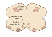 |
Figure 80: Vascular Distribution and Anatomy Relevant to the Medial Medullary Syndrome (Supplement) | Image | |
| 77 |
 |
Gaze-Evoked Nystagmus & Slow Saccades Due to Anti-GAD Antibodies in a Patient with Stiff Person Syndrome | This is a 70-year-old woman with a several year long history of imbalance and stiffness. Exam demonstrated axial and lower extremity stiffness, and ocular motor exam demonstrated gaze-evoked nystagmus (e.g., right-beating in right gaze, left-beating in left gaze, up-beating in up gaze), and mild to ... | Image/MovingImage |
| 78 |
 |
Head Movement Independent ('Sitting') Oscillopsia - A Common Symptom of Nystagmus and Saccadic Intrusions/Oscillations | 𝗢𝗿𝗶𝗴𝗶𝗻𝗮𝗹 𝗗𝗲𝘀𝗰𝗿𝗶𝗽𝘁𝗶𝗼𝗻: This video is an example of what a patient with spontaneous nystagmus or saccadic intrusions/oscillations experiences visually during the abnormal eye movements - i.e., oscillopsia (illusion of movement of the stationary ... | Image/MovingImage |
| 79 |
 |
Hemifacial Spasm | This is a 45-year-old man with intermittent left facial twitching and eyelid closure for the last 6 months. With observation, spontaneous left facial spasms were seen involving the orbicularis oculi and oris muscles. With voluntary contraction of left facial muscles, with smiling for instance, there... | Image/MovingImage |
| 80 |
 |
Ipsitorsional Quick Phases with Head Tilt in a Normal Subject | This is a demonstration of ocular counterroll, which can be seen when the head is tilted to the right or to the left. For example, when the head is tilted to the right, the top poles of both eyes should rotate toward the left ear to keep the top poles oriented with earth vertical. This is part of ... | Image/MovingImage |
| 81 |
 |
Jerk Nystagmus | 𝗢𝗿𝗶𝗴𝗶𝗻𝗮𝗹 𝗗𝗲𝘀𝗰𝗿𝗶𝗽𝘁𝗶𝗼𝗻: This is an example of jerk nystagmus due to a central vestibular lesion. The slow phase is the pathologic phase (to the left) which initiates the movement, and is followed by a fast position reset mechanism (to the right)... | Image/MovingImage |
| 82 |
 |
Latent Nystagmus and DVD in Infantile Esotropia | This is a 20-year-old woman with infantile esotropia (s/p strabismus surgery as a child) who demonstrated latent nystagmus and presumed dissociated vertical deviation (DVD) OS, which are commonly seen with infantile esotropia (also inferior oblique overaction and monocular nasotemporal asymmetry to ... | Image/MovingImage |
| 83 |
 |
Light Near Dissociation in a Tonic Pupil | 𝗢𝗿𝗶𝗴𝗶𝗻𝗮𝗹 𝗗𝗲𝘀𝗰𝗿𝗶𝗽𝘁𝗶𝗼𝗻: This is a 65-year-old woman who noticed difficulty reading and heightened sensitivity to lights OS for the last 6 months. On examination, there was mydriasis OS of about 6 mm (3 mm OD). The left (mydriatic) pupil constric... | Image/MovingImage |
| 84 |
 |
Localization of Ophthalmoplegia | 𝗢𝗿𝗶𝗴𝗶𝗻𝗮𝗹 𝗗𝗲𝘀𝗰𝗿𝗶𝗽𝘁𝗶𝗼𝗻: A table describing the localization of ophthalmoplegia. | Text |
| 85 |
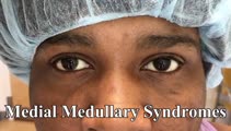 |
Medial Medullary Syndromes | This is a video of two patients who suffered small strokes involving the right medial medulla, and who presented with acute vertigo and oscillopsia. The first patient in the video had pure upbeat nystagmus, while the second patient had upbeat-torsional (towards the right ear) nystagmus in addition t... | Image/MovingImage |
| 86 |
 |
Mild 6th Nerve Palsy Due to Pontine Stroke | This is a 70-year-old woman with HTN and diabetes who presented with horizontal diplopia for several weeks, worse in right gaze. There was a very subtle abduction paresis OD with full motility elsewhere. With cover-uncover testing, there was a small esotropia in right gaze (esodeviation seen with al... | Image/MovingImage |
| 87 |
|
Organic Convergence Spasm and Nystagmus in Posterior Fossa Lymphoma | This is a 20-year-old woman, who 9 months prior to this video, first experienced episodes of vertigo and vomiting occurring when lying down or rolling over in bed. Gastrointestinal work-up was unrevealing and MRI was performed which demonstrated "multifocal nodular enhancing lesions along the ependy... | Image/MovingImage |
| 88 |
 |
Oscillopsia and Bilateral Vestibular Loss with Gentamicin Ototoxicity | Patients with bilateral vestibular loss commonly experience oscillopsia with head movements, or an inability to stabilize retinal images with subsequent bouncing or jumping of the environment due to loss of vestibular function. This causes significant blurring of vision and disorientation, dizziness... | Image/MovingImage |
| 89 |
 |
Oscillopsia: A Common Symptom of Bilateral Vestibular Loss | 𝗢𝗿𝗶𝗴𝗶𝗻𝗮𝗹 𝗗𝗲𝘀𝗰𝗿𝗶𝗽𝘁𝗶𝗼𝗻: This video is an example of what a patient with bilateral vestibular loss experiences while walking. Without a VOR, there is no mechanism to ensure retinal stability of the world with each head movement, and oscillopsia (... | Image/MovingImage |
| 90 |
 |
Paraflocculus (Tonsillar) Ocular Motor Syndrome and Dysmetria in a Chiari Malformation - Pre and Post-Operative Exams | 𝗢𝗿𝗶𝗴𝗶𝗻𝗮𝗹 𝗗𝗲𝘀𝗰𝗿𝗶𝗽𝘁𝗶𝗼𝗻: This is a 25-year-old woman presenting with 6 months or progressive imbalance, binocular vertical diplopia, and occipital headaches, which were brought on or aggravated by coughing or sneezing. Examination demonstrated hy... | Image/MovingImage |
| 91 |
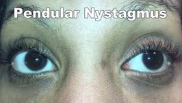 |
Pendular Nystagmus | 𝗢𝗿𝗶𝗴𝗶𝗻𝗮𝗹 𝗗𝗲𝘀𝗰𝗿𝗶𝗽𝘁𝗶𝗼𝗻: This is an example of pendular nystagmus, where like jerk nystagmus, the slow phase initiates the movement. However, unlike jerk nystagmus, there is no fast phase, but rather back to back slow phases resembling a pendulum... | Image/MovingImage |
| 92 |
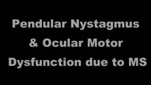 |
Pendular Nystagmus and Ocular Motor Signs in MS | 𝗢𝗿𝗶𝗴𝗶𝗻𝗮𝗹 𝗗𝗲𝘀𝗰𝗿𝗶𝗽𝘁𝗶𝗼𝗻: This is a 30-year-old man with a 15 year history of multiple sclerosis. For the last 12 months, he experienced horizontal oscillopsia. On examination, there were ocular motor abnormalities including gaze-evoked nystagmus,... | Image/MovingImage |
| 93 |
 |
Periodic Alternating Nystagmus Due to a Chiari Malformation | This patient first experienced oscillopsia 12 months prior to this video. Three months after the onset of symptoms, she was seen by neuro-ophthalmology and found to have a spontaneous, unidirectional left-beating nystagmus (that did not reverse) in addition to saccadic smooth pursuit. Oscillopsia wo... | Image/MovingImage |
| 94 |
 |
Periodic Alternating Nystagmus Due to Nodulus Stroke | This is a 70-year-old woman who experienced the acute onset of vertigo and imbalance. MRI demonstrated a diffusion-weighted imaging hyperintensity involving the nodulus (with corresponding ADC hypointensity) consistent with an acute stroke. On examination several weeks after the stroke, periodic alt... | Image/MovingImage |
| 95 |
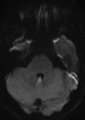 |
Periodic Alternating Nystagmus Due to Nodulus Stroke (Figure 1) | This is a 70-year-old woman who experienced the acute onset of vertigo and imbalance. MRI demonstrated a diffusion-weighted imaging hyperintensity involving the nodulus (with corresponding ADC hypointensity) consistent with an acute stroke. On examination several weeks after the stroke, periodic alt... | Image |
| 96 |
|
Positional Nystagmus During an Attack of Vestibular Migraine | 𝗢𝗿𝗶𝗴𝗶𝗻𝗮𝗹 𝗗𝗲𝘀𝗰𝗿𝗶𝗽𝘁𝗶𝗼𝗻: A 50-year-old woman presented to clinic after experiencing multiple episodes of hours-long vertigo attacks that were associated with headache, photophobia and phonophobia. She had a history of motion sickness and migraine... | Image/MovingImage |
| 97 |
 |
Posterior Canal BPPV with Fixation and with Fixation Removed | This is a 60-yo-woman with positional vertigo. In the right Dix-Hallpike position with fixation removed, there was clear upbeat-torsional nystagmus (towards the lowermost right ear) which led to the diagnosis of right posterior canal BPPV. In right Dix-Hallpike with fixation there was mainly torsion... | Image/MovingImage |
| 98 |
|
Relative Afferent Pupillary Defect in Compressive Optic Neuropathy Due to Meningioma | 𝗢𝗿𝗶𝗴𝗶𝗻𝗮𝗹 𝗗𝗲𝘀𝗰𝗿𝗶𝗽𝘁𝗶𝗼𝗻: This is a 35-year-old woman with a compressive optic neuropathy OS due to a meningioma. She had normal acuity and color OD, with 20/40 acuity and dyschromatopsia OS. There was loss of visual field OS with a mainly tempora... | Image/MovingImage |
| 99 |
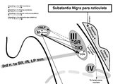 |
Sagittal Section of the Midbrain Showing Structures Related to Normal Eyelid Function | 𝗢𝗿𝗶𝗴𝗶𝗻𝗮𝗹 𝗗𝗲𝘀𝗰𝗿𝗶𝗽𝘁𝗶𝗼𝗻: During a vertical saccade, the rostral interstitial nucleus of the medial longitudinal fasciculus (riMLF) is activated, which excites the superior rectus (SR) and inferior oblique (IO) (IIIrd nerve) subnuclei. Additionall... | Image |
| 100 |
|
Sitting & Walking Oscillopsia in a Patient with Bilateral Vestibular Loss & Head Tremor | This is a 55-year-old man with oscillopsia for two reasons: He experienced oscillopsia at rest - so-called ‘sitting' oscillopsia - not from spontaneous nystagmus, but because of a combination of bilateral vestibular loss (BVL) and a mainly horizontal head tremor (this is sometimes referred to a... | Image/MovingImage |