AAO-NANOS Neuro-Ophthalmology Clinical Collection: Derived from the AAO-NANOS Clinical Neuro-Ophthalmology collection produced on CD. The images are of selected cases from the NANOS teaching slide exchange, and the CD was produced under the direction of Larry Frohman, MD and Andrew Lee, MD.
The American Academy of Ophthalmology (AAO); The North American Neuro-Ophthalmology Association (NANOS).
NOVEL: https://novel.utah.edu/
TO
| Title | Creator | Description | ||
|---|---|---|---|---|
| 76 |
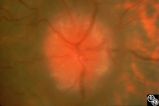 |
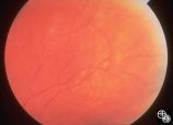
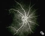
Isolated Optic Neuritis/Neuropathy | Anthony C. Arnold, MD | This 42-year-old male with pseudotumor cerebri and chronic papilledema demonstrated refractile bodies, which can been seen with chronic optic disc edema. This image demonstrates later recurrence of the refractile bodies with worsening papilledema OD. Pair with 96_01, 96_02, 96_03, 96_04, and 96_06. |
| 77 |
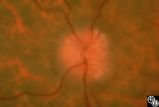 |
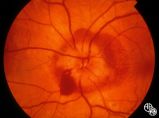
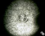
Isolated Optic Neuritis/Neuropathy | Anthony C. Arnold, MD | This 42-year-old male with pseudotumor cerebri and chronic papilledema demonstrated refractile bodies, which can be seen with chronic optic disc edema. This image demonstrates later recurrence of the refractile bodies with worsening papilledema OD. Pair with 96_01, 96_02, 96_03, 96_04, and 96_05. |
| 78 |
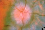 |
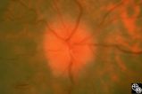
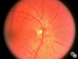
Isolated Optic Neuritis/Neuropathy | Anthony C. Arnold, MD | This 42-year-old male with pseudotumor cerebri and chronic papilledema demonstrated refractile bodies, which can be seen with chronic optic disc edema. This image shows the chronic papilledema at presentation, with associated refractile hyaline bodies at the disc periphery in both eyes. Pair with 96... |
| 79 |
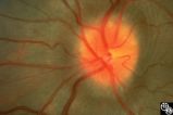 |
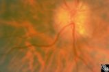
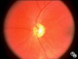
Isolated Optic Neuritis/Neuropathy | Anthony C. Arnold, MD | This image demonstrates Paton's lines in a 34-year-old patient with pseudotumor cerebri and chronic papilledema. |
| 80 |
 |
Isolated Optic Neuritis/Neuropathy | Daniel M. Jacobson MD | This 35-year-old otherwise-healthy woman developed typical optic neuritis OD with excellent recovery. She had no clinical evidence of multiple sclerosis at that time. She presented in August of 1991, at which time perivenous sheathing was seen in the retinal periphery OU. A limited workup was negati... |
| 81 |
 |
Isolated Optic Neuritis/Neuropathy | Richard H. Legge, MD | Papilledema is a term reserved for optic disc edema related to increased intracranial pressure (eg. Papilledema, sixth nerve palsy, headache), a normal neuroimaging study, and an elevated opening pressure with normal cerebrospinal fluid contents. |
| 82 |
 |
Isolated Optic Neuritis/Neuropathy | Anthony C. Arnold, MD | This 42-year-old male with pseudotumor cerebri and chronic papilledema demonstrated refractile bodies, which can been seen with chronic optic disc edema. This image exhibits decreased disc edema and resolution of the refractile bodies OD after therapy. Pair with 96_01, 96_02, 96_04, 96_05, and 96_06... |
| 83 |
 |
Isolated Optic Neuritis/Neuropathy | Anthony C. Arnold, MD | This 42-year-old male with pseudotumor cerebri and chronic papilledema demonstrated refractile bodies, which can be seen with chronic optic disc edema. This image shows the chronic papilledema at presentation, with associated refractile hyaline bodies at the disc periphery in both eyes. Pair with 96... |
| 84 |
 |
Isolated Optic Neuritis/Neuropathy | John A. Charley, MD | Several Primary Mutations result in Leber's hereditary optic neuropathy, including mitochondrial deletions at positions 11778, 3460, and 14484. Although the 11778 is the most common mutation, the 11484 has the best prognosis for spontaneous recovery. This case exhibits the 3460 mutation. |
| 85 |
 |
Isolated Optic Neuritis/Neuropathy | John A. Charley, MD | Several Primary Mutations result in Leber's hereditary optic neuropathy, including mitochondrial deletions at positions 11778, 3460, and 14484. Although the 11778 is the most common mutation, the 11484 has the best prognosis for spontaneous recovery. This case exhibits the 3460 mutation. |
| 86 |
 |
Isolated Optic Neuritis/Neuropathy | John A. Charley, MD | Several Primary Mutations result in Leber's hereditary optic neuropathy, including mitochondrial deletions at positions 11778, 3460, and 14484. Although the 11778 is the most common mutation, the 11484 has the best prognosis for spontaneous recovery. This case exhibits the 3460 mutation. |
| 87 |
 |
Isolated Optic Neuritis/Neuropathy | John A. Charley, MD | Several Primary Mutations result in Leber's hereditary optic neuropathy, including mitochondrial deletions at positions 11778, 3460, and 14484. Although the 11778 is the most common mutation, the 11484 has the best prognosis for spontaneous recovery. This case exhibits the 3460 mutation. |
| 88 |
 |
Isolated Optic Neuritis/Neuropathy | John A. Charley, MD | Several Primary Mutations result in Leber's hereditary optic neuropathy, including mitochondrial deletions at positions 11778, 3460, and 14484. Although the 11778 is the most common mutation, the 11484 has the best prognosis for spontaneous recovery. This case exhibits the 3460 mutation. |
| 89 |
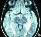 |
Magnetic Resonance Imaging in Detection of Extracranial Internal Carotid Artery Dissection | Marilyn C. Kay, MD | This 28-year-old woman presented with a 4-week history of bilateral visual loss. She had a known history of multiple sclerosis. Her vision was 20/60 OD and 20/40 OS, with an RAPD OS and optic pallor OU. Her fields and MRI are shown. Optic tract lesions usually result in an incongruous homonymous hem... |
| 90 |
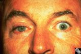 |
Migraine Syndrome | Mitchell J. Wolin, MD | The image shows a patient with cluster headache and eye displaying Horner's syndrome. |
| 91 |
 |
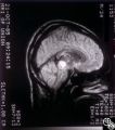
Motility Disturbances | Mitchell J. Wolin, MD | The trochlear nerve (fourth nerve) runs from the midbrain, exits dorsally, crosses in the anterior medullary velum, enters the subarachnoid space, travels within the lateral wall of the cavernous sinus, and enters the orbit through the superior orbital fissure. A trochlear nerve palsy may be due to ... |
| 92 |
 |
Motility Disturbances | Mitchell J. Wolin, MD | The trochlear nerve (fourth nerve) runs from the midbrain, exits dorsally, crosses in the anterior medullary velum, enters the subarachnoid space, travels within the lateral wall of the cavernous sinus, and enters the orbit through the superior orbital fissure. A trochlear nerve palsy may be due to ... |
| 93 |
 |
Motility Disturbances | Larry P. Frohman, MD | A 27-year-old healthy man presented for neuro-ophthalmologic evaluation for recurrent vertical binocular diplopia. In 1982, he had a minor blow to the head, and 12 days later he sustained vertical binocular diplopia, worse on downgaze, without ever having diurnal variation. His sense of taste was al... |
| 94 |
 |
Motility Disturbances | Larry P. Frohman, MD | A 27-year-old healthy man presented for neuro-ophthalmologic evaluation for recurrent vertical binocular diplopia. In 1982, he had a minor blow to the head, and 12 days later he sustained vertical binocular diplopia, worse on downgaze, without ever having diurnal variation. His sense of taste was al... |
| 95 |
 |
Motility Disturbances | Larry P. Frohman, MD | A 27-year-old healthy man presented for neuro-ophthalmologic evaluation for recurrent vertical binocular diplopia. In 1982, he had a minor blow to the head, and 12 days later he sustained vertical binocular diplopia, worse on downgaze, without ever having diurnal variation. His sense of taste was al... |
| 96 |
 |
Motility Disturbances | Larry P. Frohman, MD | A 27-year-old healthy man presented for neuro-ophthalmologic evaluation for recurrent vertical binocular diplopia. In 1982, he had a minor blow to the head, and 12 days later he sustained vertical binocular diplopia, worse on downgaze, without ever having diurnal variation. His sense of taste was al... |
| 97 |
 |
Motility Disturbances | Larry P. Frohman, MD | A 27-year-old healthy man presented for neuro-ophthalmologic evaluation for recurrent vertical binocular diplopia. In 1982, he had a minor blow to the head, and 12 days later he sustained vertical binocular diplopia, worse on downgaze, without ever having diurnal variation. His sense of taste was al... |
| 98 |
 |
Motility Disturbances | Larry P. Frohman, MD | A 27-year-old healthy man presented for neuro-ophthalmologic evaluation for recurrent vertical binocular diplopia. In 1982, he had a minor blow to the head, and 12 days later he sustained vertical binocular diplopia, worse on downgaze, without ever having diurnal variation. His sense of taste was al... |
| 99 |
 |
Motility Disturbances | Larry P. Frohman, MD | This 5-year-old child presented with a 70 PD exotropia OS and a right face turn. She had a CT scan of the head at age 4 months that was normal , and she was felt to have an isolated left medial rectus paresis. Her acuity was 20/20 OU. She could fuse with a large face turn, and was orthomorphic is ex... |
| 100 |
 |
Motility Disturbances | Larry P. Frohman, MD | This 5-year-old child presented with a 70 PD exotropia OS and a right face turn. She had a CT scan of the head at age 4 months that was normal , and she was felt to have an isolated left medial rectus paresis. Her acuity was 20/20 OU. She could fuse with a large face turn, and was orthomorphic is ex... |