Collection of materials relating to neuro-ophthalmology as part of the Neuro-Ophthalmology Virtual Education Library.
NOVEL: https://novel.utah.edu/
TO
- NOVEL233
| Title | Creator | Description | Subject | ||
|---|---|---|---|---|---|
| 76 |
 |
Lacrimal Pathways: Anatomy and Physiology | Sari Yordi, MD | Video lecture covering anatomy and physiology of the lacrimal pathways. | Lacrimal Pathways |
| 77 |
 |
Aqueous and Vitreous Humor | Sari Yordi, MD | Narrated lecture on the aqueous and vitreous humor. | Aqueous; Vitreous Humor |
| 78 |
 |
Physiology of Intraocular Pressure | Aakash Patel; Amanda Henderson, MD | Overview of the physiology of intraocular pressure. | Intraocular Pressure; Physiology |
| 79 |
 |
Optic Chiasm | Yesha Shah, BSA, BBA; Amanda Henderson, MD | Overview of the anatomy of the optic chiasm. | Optic Chiasm; Anatomy |
| 80 |
 |
Control of the Eye Movements: Neuroanatomy Video Lab - Brain Dissections | Suzanne S. Stensaas, PhD | Disturbances in eye movements can provide important clues for localization of neurological damage. The role of the frontal eye fields in horizontal gaze is stressed. The need to coordinate cranial nerves on both sides of the brain stem introduces the medial longitudinal fasciculus and its role in co... | Medial Longitudinal Fasciculus; Third Cranial Nerve; Sixth Cranial Nerve; Internuclear Ophthalmoplegia; Nystagmus; Brain; Dissection |
| 81 |
 |
Optic Nerve Sheath Fenestration | Raed Behbehani, MD | Optic nerve sheath fenestration is performed to manage papilledema causing progressive loss of vision , due to raised intracranial pressure from Idiopathic Intracranial Hypertension or Cerebral Venous Sinus Thrombosis. The procedure is usually performed in cases of severe visual field loss or when m... | Optic Nerve Sheath Fenestration |
| 82 |
 |
Superonasal Transconjunctival Optic Nerve Sheath Decompression (stONSD) | Kevin E. Lai, MD; Kenneth C. Lao, MD; Peter L. Hildebrand, MD; Bradley K. Farris, MD | This video demonstrates the surgical technique and outcomes of a modified medial transconjunctival approach to optic nerve sheath decompression (ONSD). Disease/Diagnosis: Papilledema; Idiopathic Intracranial Hypertension (IIH). | Superonasal Transconjunctival Optic Nerve Sheath Decompression (ONSD); Surgical Technique |
| 83 |
 |
Lateral Orbitotomy with Bone Window | Richard C. Allen, MD, PhD, FACS | Narrated video of lateral orbitotomy with bone removal for improved orbital decompression. | Orbitotomy; Orbital Decompression |
| 84 |
 |
Lateral Orbitotomy #3 | Richard C. Allen, MD, PhD, FACS | Narrated video of lateral orbitotomy and biopsy of presumed benign cavernous hemangioma. | Orbital Biopsy; Orbitotomy |
| 85 |
 |
Lateral Orbitotomy #2 | Richard C. Allen, MD, PhD, FACS | Narrated video of lateral orbitotomy and biopsy of presumed orbital inflammatory condition. | Orbital Biopsy; Orbitotomy |
| 86 |
 |
Comparison of the Motor Systems: Neuroanatomy Video Lab - Brain Dissections | Suzanne S. Stensaas, PhD | A comparison of the three major motor systems focuses on categorizing motor problems as corticospinal tract, cerebellar, or basal ganglia. It begins with 12 minutes of gross anatomical structures and pathway review followed by clinical video clips demonstrating clinical features of disease of each o... | Corticospinal Tract; Cerebellar; Basal Ganglia; Motor Systems; Brain; Dissection |
| 87 |
 |
Basal Ganglia: Neuroanatomy Video Lab - Brain Dissections | Suzanne S. Stensaas, PhD | Structures involved in involuntary movements are shown on models, in animations, and on gross coronal and axial sections. Diagrams show the cortex-to-cortex loop and the nigrostriatal pathway. The direct and indirect pathways are shown. The direct pathway facilitates movement while the indirect path... | Basal Ganglia; Involuntary Movements; Brain; Dissection |
| 88 |
 |
Cortical Localization: Neuroanatomy Video Lab - Brain Dissections | Suzanne S. Stensaas, PhD | The lobes of the brain are defined together with their major functions. The visual field representation in the occipital lobe is explained with a diagram. Speech areas and the major types of aphasia are discussed in the dominant hemisphere and parietal lesions of neglect and spatial orientation are ... | Cortical Localization; Brain; Dissections |
| 89 |
 |
Introduction: Neuroanatomy Video Lab - Brain Dissections | Suzanne S. Stensaas, PhD | The regions and lobes of the brain are identified along with some of the nerves and vessels. The basic functions of the cortex of each lobe are introduced along with principal sulci and gyri. The importance of the left hemisphere for language and the temporal lobe in memory are mentioned along with ... | Brain; Dissections |
| 90 |
 |
Brain Stem & Reflexes: Neuroanatomy Video Lab - Brain Dissections | Suzanne S. Stensaas, PhD | The cranial nerves are reviewed again on a specimen with vessels. Next, landmarks on gross brain stem sections are shown. Stressed are the three reflexes associated with each of the three levels: pupillary, corneal and gag reflexes and their associated cranial nerves. Finally cross sections of myeli... | Brain Stem; Reflexes |
| 91 |
 |
Cerebral Circulation: Neuroanatomy Video Lab - Brain Dissections | Suzanne S. Stensaas, PhD | The major vessels of the anterior and posterior circulation are demonstrated along with the Circle of Willis on both a model and in an animation. The distribution of the three major cerebral arteries is demonstrated along with the concept of a watershed zone. A gross specimen with good vessels is al... | Cerebral Circulation; Brain; Dissections |
| 92 |
 |
Sixth nerve palsy or not? (Video) | Vivian Paraskevi Douglas; Konstantinos Douglas; Nurhan Torun | Here in we present a case of a 72-yearold Caucasian female with remote history of breast cancer who presented with diplopia in the right gaze over the past several months. There had been no improvement after a 5-day course of steroids by an outside ophthalmologist. On neuro-ophthalmic examination, t... | Abduction deficit; Diplopia; Orbital imaging |
| 93 |
 |
Cerebellum: Neuroanatomy Video Lab - Brain Dissections | Suzanne S. Stensaas, PhD | The gross features of the cerebellum are shown. The three peduncles are demonstrated, noting their input and output to and from the cerebellum. Emphasis is given to symptoms of cerebellar disease appearing on the same side of the body. Special emphasis is given to cortical-cerebellar connections, st... | Cerebellum; Cortical-Cerebellar Connection; Brain; Dissection |
| 94 |
 |
Control of the Pupil: Neuroanatomy Video Lab - Brain Dissections | Suzanne S. Stensaas, PhD | Through diagrams, animations and gross specimens the constriction and dilation of the pupil by the autonomic nervous system are described. Both the parasympathetic and sympathetic control are traced and the importance of a constricted pupil, Horner's Syndrome, and temporal lobe (uncal) herniation (d... | Brain; Dissections |
| 95 |
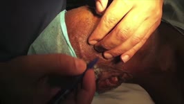 |
Temporal Artery Biopsy | Raed Behbehani, MD | This is a video of Superficial Temporal Artery Biopsy done under local anaesthesia for a patient who was suspected to have Giant Cell Arteritis (GCA. GCA is vasculitis of the medium sized vessels than can lead to permanent visual loss by causing Arteritis Ischemic Optic Neuropathy. The diagnosis of ... | Temporal Artery Biopsy; Giant Cell Arteritis |
| 96 |
 |
Principles of Strabismus Surgery | Michael B. Yang, MD | A video demonstrating a medial rectus recession. | Strabismus; Surgery; Surgery Demonstrations |
| 97 |
 |
Arteriovenous Malformation | Justin Gibson, MD; Charles Prestigiacomo, MD | A diagnostic cerebroangiogram performed on a patient who presented with worst headache of life, found to have a Fisher Grade 3 subarachnoid hemorrhage. | Angiogram; Arteriovenous Malformation; AVM |
| 98 |
 |
Large Vessel Occlusion | Justin Gibson, MD; Charles Prestigiacomo, MD | Example of a diagnostic cerebroangiogram performed on a patient undergoing a stroke. | Angiogram; Stroke |
| 99 |
 |
Cerebral Aneurysm | Justin Gibson, MD; Charles Prestigiacomo, MD | Cerebral angiogram of a patient with an arteriovenous malformation, or AVM. | Angiogram; Cerebral Aneurysm |
| 100 |
 |
Normal Angiogram | Justin Gibson, MD; Charles Prestigiacomo, MD | Example of a normal diagnostic cerebroangiogram. | Angiogram |