A collection of videos relating to the diagnosis and treatment of eye movement disorders. This collection includes many demonstrations of examination techniques.
Dan Gold, D.O., Associate Professor of Neurology, Ophthalmology, Neurosurgery, Otolaryngology - Head & Neck Surgery, Emergency Medicine, and Medicine, The Johns Hopkins School of Medicine.
A collection of videos relating to the diagnosis and treatment of eye movement disorders.
NOVEL: https://novel.utah.edu/
TO
| Title | Description | Type | ||
|---|---|---|---|---|
| 51 |
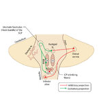 |
Figure 50: Anatomy and Physiology of the Saccadic Pathways | When a saccade is desired (or reflexively triggered), signals project from the saccade-related cortical eye fields to the superior colliculus, which serves to integrate and relay commands to the saccade generating brainstem circuitry. The inferior cerebellar peduncle (ICP) carries climbing fibers to... | Image |
| 52 |
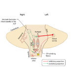 |
Figure 51: Lateral Medullary Lesion Causing Saccadic Dysmetria | A lesion of the left lateral medulla and inferior cerebellar peduncle (ICP) will cause decreased climbing fiber inhibition of the left dorsal vermis causing simple-spike (inhibitory) discharge of Purkinje cells to increase. Increased Purkinje cell firing leads to increased inhibition of the ipsilate... | Image |
| 53 |
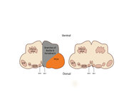 |
Figure 53: Vascular Distribution and Anatomy Relevant to the Lateral Medullary (Wallenberg) Syndrome | This axial section of the medulla highlights those structures that, when damaged, are responsible for the vestibular and ocular motor features of the Wallenberg syndrome. The nucleus prepositus hypoglossi (NPH) and medial vestibular nucleus (MVN) complex is important for horizontal gaze-holding (neu... | Image |
| 54 |
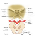 |
Figure 64: The Course of the 3rd (III) Nerve | The 3rd nucleus lies at the ventral border of the periaqueductal gray matter, at the level of the superior colliculus. In between the two nuclei is the midline central caudal nucleus (CCN), which innervates bilateral levator palpebrae muscles (explaining how a unilateral nuclear 3rd can cause bilate... | Image |
| 55 |
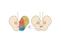 |
Figure 65: Vascular Distribution and Anatomy (Including 3rd Nerve) of the Rostral Midbrain | In this axial section of the midbrain at the level of the superior colliculus, the paired 3rd nuclei are located ventral to the periaqueductal grey, and the midline central caudal nucleus (CCN) is located in between. The fascicles that exit the IIIrd nuclei carry the fibers destined to innervate the... | Image |
| 56 |
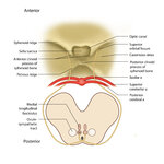 |
Figure 68: The Course of the 4th (IV) Nerve | The 4th nucleus lies at the ventral border of the periaqueductal gray matter, at the level of the inferior colliculus. The fascicles exit the nucleus dorsally and decussate at the anterior medullary velum (anterior floor of the fourth ventricle), and then exit the brainstem dorsally. The peripheral ... | Image |
| 57 |
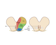 |
Figure 69: Vascular Distribution and Anatomy (Including 4th Nerve) of the Caudal Midbrain | In this axial section of the midbrain at the level of the inferior colliculus, the 4th nuclei are located ventral to the periaqueductal grey, dorsal to the medial longitudinal fasciculus (MLF) and medial to the oculosympathetic tract. Fascicles exit the nucleus dorsally and decussate at the anterior... | Image |
| 58 |
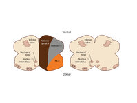 |
Figure 80: Vascular Distribution and Anatomy Relevant to the Medial Medullary Syndrome | This axial section of the medulla highlights those structures that, when damaged, are often responsible for spontaneous upbeat nystagmus (UBN). The nucleus of Roller and nucleus intercalatus normally have an inhibitory influence over the cerebellar flocculus, and when there is a lesion of Roller/int... | Image |
| 59 |
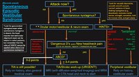 |
An Approach to the Patient with (Recent Onset) Spontaneous Episodic Vestibular Syndrome | 𝗢𝗿𝗶𝗴𝗶𝗻𝗮𝗹 𝗗𝗲𝘀𝗰𝗿𝗶𝗽𝘁𝗶𝗼𝗻: A vascular etiology should always be on the differential diagnosis of the recent onset of the spontaneous (unprovoked) episodic vestibular syndrome (EVS), especially in the older population and when vascular risk factors ... | Image |
| 60 |
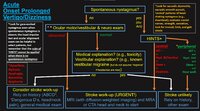 |
An Approach to the Patient with Acute Onset Prolonged Vertigo | 𝗢𝗿𝗶𝗴𝗶𝗻𝗮𝗹 𝗗𝗲𝘀𝗰𝗿𝗶𝗽𝘁𝗶𝗼𝗻: A vascular etiology should always be on the differential diagnosis of the acute onset prolonged vertigo, especially in the older population and when vascular risk factors are present. However, young patients can suffer fr... | Image |