A collection of videos relating to the diagnosis and treatment of eye movement disorders. This collection includes many demonstrations of examination techniques.
Dan Gold, D.O., Associate Professor of Neurology, Ophthalmology, Neurosurgery, Otolaryngology - Head & Neck Surgery, Emergency Medicine, and Medicine, The Johns Hopkins School of Medicine.
A collection of videos relating to the diagnosis and treatment of eye movement disorders.
NOVEL: https://novel.utah.edu/
TO
| Title | Description | Type | ||
|---|---|---|---|---|
| 51 |
 |
Sagittal Section of the Brainstem Showing Structures Related to Normal Eyelid Function | Seen here is a sagittal view of the brainstem, with the structures relevant to normal eyelid function highlighted. The M-group, which can be found medial to the riMLF (coordinates eye and lid movements), has (weak) projections to the facial nucleus for frontalis muscle contraction, and (strong) proj... | Image |
| 52 |
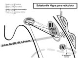 |
Sagittal Section of the Midbrain Showing Structures Related to Normal Eyelid Function | 𝗢𝗿𝗶𝗴𝗶𝗻𝗮𝗹 𝗗𝗲𝘀𝗰𝗿𝗶𝗽𝘁𝗶𝗼𝗻: During a vertical saccade, the rostral interstitial nucleus of the medial longitudinal fasciculus (riMLF) is activated, which excites the superior rectus (SR) and inferior oblique (IO) (IIIrd nerve) subnuclei. Additionall... | Image |
| 53 |
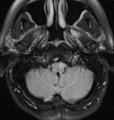 |
Subtle Torsional Pendular Nystagmus in Oculopalatal Tremor (OPT) (Figure 1) | This is a 50-year-old woman who presented with imbalance, and MRI demonstrated a right cerebellar cavernous malformation. She underwent surgery to resect the malformation, and post-operatively experienced right hemiparesis and ataxia. Six months after the surgery, balance worsened and vision became ... | Image |
| 54 |
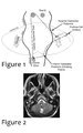 |
Triangle of Guillain-Mollaret | Seen here is a schematic representation of the Gullain-Mollaret triangle (Figure 1), also referred to as the dentato-olivary pathway, reflecting the 3 points of this imaginary triangle - 1) dentate nucleus, 2) red nucleus, and 3) inferior olivary nucleus. The olive sends decussating climbing fibers ... | Image |
| 55 |
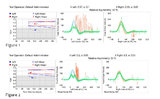 |
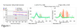
Using Video Head Impulse Testing to Unmask Covert Saccades in Compensated Vestibular Neuritis (Figures 1 and 2) | This is a 30-year-old woman who experienced the acute vestibular syndrome (prolonged vertigo for >24 hours, nausea, unsteadiness, spontaneous nystagmus, head motion intolerance) and was diagnosed with vestibular neuritis. This diagnosis was based on a positive head impulse test to the left (see Figu... | Image |
| 56 |
 |
The Utriculo-Ocular Motor Pathways - Physiologic and Pathologic Ocular Tilt Reaction: OTR Diagram Pathologic EOMs Labelled (Figure 3) | A skew deviation is a non-paralytic vertical ocular misalignment that is due to imbalance in the utriculo-ocular motor pathways. While vestibular jerk nystagmus is a consequence of static semicircular canal pathway imbalance (e.g., left-beating nystagmus due to acute right vestibular hypofunction fr... | Image |
| 57 |
 |
The Utriculo-Ocular Motor Pathways - Physiologic and Pathologic Ocular Tilt Reaction: Pathologic OTR (Figure 2) | A skew deviation is a non-paralytic vertical ocular misalignment that is due to imbalance in the utriculo-ocular motor pathways. While vestibular jerk nystagmus is a consequence of static semicircular canal pathway imbalance (e.g., left-beating nystagmus due to acute right vestibular hypofunction fr... | Image |
| 58 |
 |
The Utriculo-Ocular Motor Pathways - Physiologic and Pathologic Ocular Tilt Reaction: Physiologic Ocular Tilt Reaction (OTR) (Figure 1) | A skew deviation is a non-paralytic vertical ocular misalignment that is due to imbalance in the utriculo-ocular motor pathways. While vestibular jerk nystagmus is a consequence of static semicircular canal pathway imbalance (e.g., left-beating nystagmus due to acute right vestibular hypofunction fr... | Image |
| 59 |
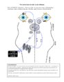 |
Vertical Semicircular Canal Pathways | Anterior Canal Pathway Afferents that originate in the anterior canals (AC) of the peripheral labyrinth first synapse in the ipsilateral vestibular nucleus. Three pathways exist: 1) medial longitudinal fasciculus (MLF) - right AC afferents to right medial vestibular nucleus (MVN), decussate and asc... | Image |
| 60 |
 |
Vestibular Neuritis with + Head Impulse Test and Unidirectional Nystagmus (Figure 1) | Vestibular neuritis is the most common cause of the acute vestibular syndrome, which is characterized by continuous vertigo and spontaneous nystagmus lasting days. It may be mimicked by central causes, including stroke, but in the hands of subspecialists, the HINTS+ (Head Impulse, Nystagmus, Test of... | Image |