Best known for his world-renowned neuro-ophthalmology unit based at the University of California, San Francisco, William Hoyt, MD collected here more than 850 of his best images covering a wide range of disorders.
William F. Hoyt, MD, Professor Emeritus of Ophthalmology, Neurology and Neurosurgery, Department of Ophthalmology, University of California, San Francisco.
NOVEL: https://novel.utah.edu/
TO
Filters: Collection: "ehsl_novel_wfh"
| Title | Description | Type | ||
|---|---|---|---|---|
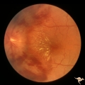
| 51 |
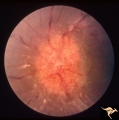 |
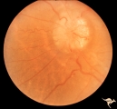
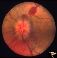
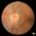
Chronic Papilledema in Resolution. Sequence | Left eye at presentation. Chronic papilledema. Anatomy: Optic disc Pathology: Papilledema Disease/Diagnosis: Papilledema | Image |
| 52 |
 |
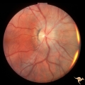
Chronic Papilledema in Resolution. Sequence | Left eye 2 weeks after presentation. Chronic papilledema in resolution. Note first evidence of a vertical choroidal fold. Anatomy: Optic disc. Pathology: Papilledema. Disease/Diagnosis: Papilledema. | Image |
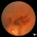
| 53 |
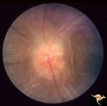 |
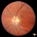
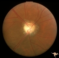
Chronic Papilledema in Resolution. Sequence | Left eye 4 weeks after presentation. Chronic papilledema in resolution. Notice more extensive vertical choroidal fold temporally ("high-water" marks) | Image |
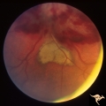
| 54 |
 |
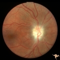
Chronic Papilledema in Resolution. Sequence | Left eye 7 weeks after presentation. Chronic papilledema in resolution. Note the profound optic atrophy with blurred disc margins and circumferential receptor layer folds ("high-water" marks) | Image |
| 55 |
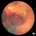 |
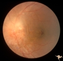
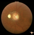
Chronic Papilledema with Hemorrhagic and Exudative Complications | Left eye at presesntation. Chronic papilledema with hemorrhagic and exudative complications due to Pseudotumor cerebri. Anatomy: Optic disc Pathology: Papilledema Disease/Diagnosis: Chronic papilledema with hemorrhagic and exudative complications. | Image |
| 56 |
 |
Chronic Papilledema with Hemorrhagic and Exudative Complications | Left eye one month after presentation. Resolving hemorrhage. Chronic papilledema with hemorrhagic and exudative complications due to Pseudotumor cerebri. Anatomy: Optic disc. Pathology: Papilledema. Disease/Diagnosis: Chronic papilledema with hemorrhagic and exudative complications | Image |
| 57 |
 |
Chronic Papilledema with Hemorrhagic and Exudative Complications | Left eye one month after presentation. View below of resolving subretinal hemorrhage. Chronic papilledema with hemorrhagic and exudative complications due to Pseudotumor cerebri. | Image |
| 58 |
 |
Chronic Papilledema with Hemorrhagic and Exudative Complications | Left eye one month after presentation. View above of resolving preretinal hemorrhage. Chronic papilledema with hemorrhagic and exudative complications due to Pseudotumor cerebri. | Image |
| 59 |
 |
Chronic Papilledema with Pseudo Drusen | Right eye. Meningioma. Pseudo drusen from chronic papilledema. Woman. Anatomy: Optic disc Pathology: Papilledema Disease/Diagnosis: Chronic papilledema with pseudo drusen | Image |
| 60 |
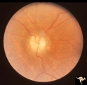 |
Chronic Papilledema with Pseudo Drusen | Left eye. Meningioma. Pseudo drusen from chronic papilledema. The patient's meningioma had blinded her left eye and caused chronic elevated intracranial pressure. Woman. Anatomy: Optic disc Pathology: Papilledema Disease/Diagnosis: Chronic papilledema with pseudo drusen | Image |
| 61 |
 |
Chronic Papilledema with Pseudo Drusen | Left eye of 51 year old, 220 pound black woman. Pseudotumor cerebri, pseudo drusen, exudates. Anatomy: Optic disc. Pathology: Papilledema Disease/Diagnosis: Chronic papilledema with pseudo drusen | Image |
| 62 |
 |
Chronic Papilledema with Pseudo Drusen | Chronic papilledema with pseudo drusen. Residual choroidal folds. Pseudo drusen. Anatomy: Optic disc. Pathology: Papilledema. Disease/Diagnosis: Chronic papilledema with pseudo drusen. | Image |
| 63 |
 |
Chronic Papilledema with Pseudo Drusen | Right eye. Chronic papilledema with pseudo drusen due to cerebral pontine angle tumor. Anatomy: Optic disc Pathology: Papilledema Disease/Diagnosis: Chronic papilledema with pseudo drusen | Image |
| 64 |
 |
Chronic Papilledema with Pseudo Drusen | Left eye. Chronic papilledema with pseudo drusen due to cerebral pontine angle tumor. Anatomy: Optic disc. Pathology: Papilledema Disease/Diagnosis: Chronic papilledema with pseudo drusen. | Image |
| 65 |
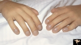 |
Cyanotic Heart Disease with Clubbing of Fingernails | Note the cyanotic nail beds and clubbing. Anatomy: Optic disc. Pathology: Papilledema. Disease/Diagnosis: Pseudotumor due to cyanotic heart disease. Clinical: Young boy with clubbing. | Image |
| 66 |
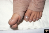 |
Cyanotic Heart Disease with Clubbing of Toes | Bilateral Papilledema with cyanotic heart disease. Anatomy: Optic disc. Pathology: Papilledema. Disease/Diagnosis: Pseudotumor due to cyanotic heart disease. Clinical: Young boy with clubbing. | Image |
| 67 |
 |
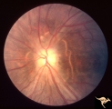
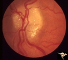
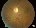
Early Papilledema due to Brain Tumor - Resolving | Left eye. Same eye as P_34a. One month post op, papilledema resolving. Boy. Anatomy: Optic disc. Pathology: Papilledema. Disease/Diagnosis: Papilledema from posterior fossa hemangioblastoma. | Image |
| 68 |
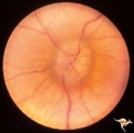
 |
Early Papilledema due to Tumor | Left eye. Asymmetric Papilledema with posterior fossa hemangioblastoma. Left - moderate papilledema. Blurring of disc. Young man. Anatomy: Optic disc. Pathology: Papilledema. Disease/Diagnosis: Papilledema from posterior fossa hemangioblastoma. | Image |
| 69 |
 |
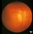
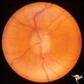
ID01 Post Papilledema Gliosis | Post papilledema milky gliosis with arteriolar constriction, 1982, right eye, pair with ID_2. Anatomy: Optic disc. Pathology: Post papilledema atrophy and gliosis due to huge anterior communicating artery aneurysm. Disease/ Diagnosis: Elevated intracranial pressure from aneurysm. Clinical: Diminishe... | Image |
| 70 |
 |
ID02 Post Papilledema Gliosis | Post papilledema milky gliosis with arteriolar constriction and atrophy, 1982, left eye, pair with ID_1. Anatomy: Optic disc. Pathology: Post papilledema atrophy and gliosis due to huge anterior communicating artery aneurysm. Disease/ Diagnosis: Elevated intracranial pressure from aneurysm. Clinical... | Image |
| 71 |
 |
ID03a Post Papilledema Atrophy with Marked Gliosis | Post papilledema atrophy with marked gliosis in a patient with pseudotumor cerebri. Patient weighed over 300 pounds. Right eye blind. 1981. Right eye. Pair with ID_3b. Anatomy: Optic disc. Pathology: Post papilledema atrophy and gliosis from long standing elevated intracranial pressure. Disease/ Dia... | Image |
| 72 |
 |
ID03b Post Papilledema Atrophy with Marked Gliosis | Post papilledema atrophy with marked gliosis in a patient with pseudotumor cerebri. Patient weighed over 300 pounds. Left eye has visual field defects. 1981, right eye, pair with ID_3a. Anatomy: Optic disc. Pathology: Post papilledema atrophy and gliosis from long standing elevated intracranial pres... | Image |
| 73 |
 |
ID04a Post Papilledema Atrophy with Marked Gliosis | Post papilledema atrophy with marked gliosis in a patient with pseudotumor cerebri, 1985, right eye, pair with ID_4b, Note "high water" marks in peripapillary pigment epithelial layer. Anatomy: Optic disc. Pathology: Post papilledema atrophy and gliosis from long standing elevated intracranial press... | Image |
| 74 |
 |
ID04b Post Papilledema Atrophy with Marked Gliosis | Post papilledema atrophy with marked gliosis in a patient with pseudotumor. Nasal ovoid absence of the retinal pigment epithelium. Presumably a defect from the long standing papilledema. 1985,. Right eye, pair with ID_4a. Anatomy: Optic disc. Pathology: Post papilledema atrophy and gliosis from long... | Image |
| 75 |
 |
ID05a Post Papilledema Optic Atrophy from Pseudotumor Cerebri | Left eye, October 1999, Post papilledema optic atrophy from pseudotumor cerebri. Note optociliary veins in both discs. Gliosis and partial pallor following long standing papilledema and intracranial pressure. Anatomy: Optic disc. Pathology: Post papilledema atrophy and gliosis from long standing el... | Image |