Best known for his world-renowned neuro-ophthalmology unit based at the University of California, San Francisco, William Hoyt, MD collected here more than 850 of his best images covering a wide range of disorders.
William F. Hoyt, MD, Professor Emeritus of Ophthalmology, Neurology and Neurosurgery, Department of Ophthalmology, University of California, San Francisco.
NOVEL: https://novel.utah.edu/
TO
Filters: Collection: "ehsl_novel_wfh"
| Title | Description | Type | ||
|---|---|---|---|---|
| 51 |
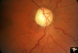 |
IE15a End Stage Leber Optic Neuropathy | End stage Leber's Optic Neuropathy. Severe diffuse pallor. Right eye. Pair with 15b. Anatomy: Optic disc. Pathology: Optic neuropathy. Disease/ Diagnosis: Leber's optic neuropathy. Clinical: Blindness. | Image |
| 52 |
 |
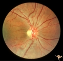
IE03 Acute Leber Optic Neuropathy | Acute stage of Leber optic neuropathy with microangiopathy and peripapillary nerve fiber layer thickening. The temporal nerve fiber layer is already showing atrophy. Central vision is grossly reduced. 1971. Anatomy: Optic disc. Pathology: Optic neuropathy. Disease/ Diagnosis: Leber's optic neuropath... | Image |
| 53 |
 |
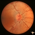
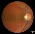
IE06 Subacute Leber Optic Neuropathy | Subacute Leber's optic neuropathy with microangiopathy with distinct temporal disc pallor. 1971. Anatomy: Optic disc. Pathology: Optic neuropathy. Disease/ Diagnosis: Leber's optic neuropathy. Clinical: Large central vision loss. | Image |
| 54 |
 |
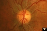
IE07 Subacute Leber Optic Neuropathy | Subacute Leber's optic neuropathy with microangiopathy. 1973. Anatomy: Optic disc. Pathology: Optic neuropathy. Disease/ Diagnosis: Leber's optic neuropathy. Clinical: Early central vision loss. | Image |
| 55 |
 |
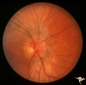
IE08a Subacute Leber Optic Neuropathy | Subacute Leber Optic Neuropathy with temporal atrophy. August 5, 1980. Pair with IE_1, 2a&b, IE_8b, IE_9a&b. Anatomy: Optic disc. Pathology: Optic neuropathy. Disease/ Diagnosis: Leber's optic neuropathy. Clinical: Visual loss. | Image |
| 56 |
 |
IE08b Subacute Leber Optic Neuropathy | Subacute Leber Optic Neuropathy with temporal atrophy. August,1980. Pair with IE_1, 2a&b, IE_8a, IE_9a&b. Anatomy: Optic disc. Pathology: Optic neuropathy. Disease/ Diagnosis: Leber's optic neuropathy. Clinical: Visual loss. | Image |
| 57 |
 |
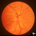
IE09a Chronic Leber Optic Neuropathy | Chronic Leber Optic Neuropathy with advancing temporal pallor. Notice the nerve fiber layer thickening has diminished. November 13, 1980. Pair with IE_1, 2a&b, IE_9b, IE_8a&b. Anatomy: Optic disc. Pathology: Optic neuropathy. Disease/ Diagnosis: Leber's optic neuropathy. Clinical: Blindness. | Image |
| 58 |
 |
IE09b Chronic Leber Optic Neuropathy | Chronic Leber Optic Neuropathy with advancing temporal pallor. Notice the nerve fiber layer thickening has diminished. November 13, 1980. Pair with IE_1, 2a&b, IE_9a, IE_8a&b. Anatomy: Optic disc. Pathology: Optic neuropathy. Disease/ Diagnosis: Leber's optic neuropathy. Clinical: Blindness. | Image |
| 59 |
 |
IE10a Chronic Leber Optic Neuropathy | Chronic Leber's Optic Neuropathy, August 8, 1969. Anatomy: Optic disc. Pathology: Optic neuropathy. Disease/ Diagnosis: Leber's optic neuropathy. Clinical: Blindness. | Image |
| 60 |
 |
IE10b Subacute Leber Optic Neuropathy | Subacute Leber's Optic Neuropathy, August 8, 1969, Left eye, pair with IE_10a, c. Anatomy: Optic disc. Pathology: Optic neuropathy. Disease/ Diagnosis: Leber's optic neuropathy. Clinical: Blindness. | Image |
| 61 |
 |
IE10c Chronic Leber Optic Neuropathy | February 12, 1970, Chronic Leber's Optic Neuropathy, 6 month follow up from 10b. Thickening of the nerve fiber layer is gone. Left eye, pair with IE_10a, c. Anatomy: Optic disc. Pathology: Optic neuropathy. Disease/ Diagnosis: Leber's optic neuropathy. Clinical: Blindness. | Image |
| 62 |
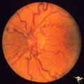 |
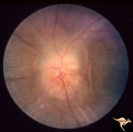
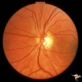
Bilateral Papilledema from Non-tumor Etiology | Bilateral Papilledema with tortuous dilated veins from chronic lung disease with cyanosis. Note the remarkable tortuosity of retinal veins, evidence of retinal cyanosis. Anatomy: Optic disc. Pathology: Bilateral papilledema. Disease/Diagnosis: Pseudotumor due to chronic lung disease. Clinical notes:... | Image |
| 63 |
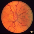 |
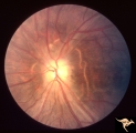
Bilateral Papilledema from Non-tumor Etiology | Right eye. Bilateral Papilledema with tortuous dilated veins from chronic lung disease with cyanosis. Note the remarkable toruosity of retinal veins, evidence of retinal cyanosis. Anatomy: Optic disc. Pathology: Bilateral papiledema. Disease/Diagnosis: Pseudotumor due to chronic lung disease. Clinic... | Image |
| 64 |
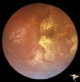 |
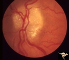
Bilateral Papilledema with Exudative Retinopathy | Right eye. Bilateral Papilledema with exudative retinopathy from vitamin A toxicity. Young boy. Near blind. Anatomy: Optic disc; Retina. Pathology: Bilateral papilledema; exudative retinopathy. Disease/Diagnosis: Hypervitaminosis A causing blindness. Clinical notes: Nearly blind; Headache. | Image |
| 65 |
 |
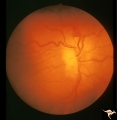
Bilateral Papilledema with Exudative Retinopathy | Left eye. Bilateral Papilledema with exudative retinopathy from vitamin A toxicity. Young boy. Near blind. Anatomy: Optic disc; Retina. Pathology: Bilateral papilledema; exudative retinopathy. Disease/Diagnosis: Hypervitaminosis A causing blindness. Clinical notes: Nearly blind; Headache. | Image |
| 66 |
 |
Bilateral Papilledema with Exudative Retinopathy | Left eye. Bilateral Papilledema with exudative retinopathy from vitamin A toxicity. Young boy. Near blind. Anatomy: Optic disc; Retina. Pathology: Bilateral papilledema; exudative retinopathy. Disease/Diagnosis: Hypervitaminosis A causing blindness. Clinical notes: Nearly blind; Headache. | Image |
| 67 |
 |
C12 Morning Glory Disc | "Morning Glory" disc. Patient 11 year old. Anatomy: Optic disc. | Image |
| 68 |
 |
C34 Anomalous Pale Disc | Multiple cilioretinal arteries. Anomalous venous exit from nasal edge of optic disc (Vein of Kraupa). Visual function normal. Anatomy: Optic disc. | Image |
| 69 |
 |
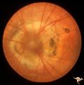
Chronic Papilledema in Resolution. Sequence | Left eye 4 weeks after presentation. Chronic papilledema in resolution. Notice more extensive vertical choroidal fold temporally ("high-water" marks) | Image |
| 70 |
 |
Chronic Papilledema in Resolution. Sequence | Left eye 7 weeks after presentation. Chronic papilledema in resolution. Note the profound optic atrophy with blurred disc margins and circumferential receptor layer folds ("high-water" marks) | Image |
| 71 |
 |
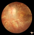
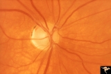
Chronic Papilledema with Pseudo Drusen | Left eye of 51 year old, 220 pound black woman. Pseudotumor cerebri, pseudo drusen, exudates. Anatomy: Optic disc. Pathology: Papilledema Disease/Diagnosis: Chronic papilledema with pseudo drusen | Image |
| 72 |
 |
Chronic Papilledema with Pseudo Drusen | Chronic papilledema with pseudo drusen. Residual choroidal folds. Pseudo drusen. Anatomy: Optic disc. Pathology: Papilledema. Disease/Diagnosis: Chronic papilledema with pseudo drusen. | Image |
| 73 |
 |
Diffuse Atrophy | Primary optic atrophy following optic neuritis. 1960. Note absence of all retinal nerve fiber layer reflex in the peripapillary retina. The retinal vessels appear to lie on the retina without any tissue surrounding them. Normal looking arterioles. Anatomy: Optic disc. Pathology: Optic atrophy. Disea... | Image |
| 74 |
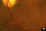 |
Diffuse Atrophy | Primary optic atrophy from optic nerve compression by aneurysm. Note narrowing of retinal arterioles. Close up showing arcuate streaks of nerve fibers entering inferior optic disc. Pair with IIA1_7. Anatomy: Optic disc. Pathology: Optic atrophy. Disease/Diagnosis: Optic atrophy due to giant aneurysm... | Image |
| 75 |
 |
Diffuse Atrophy | Primary optic atrophy following head injury. 1982. Normal looking arterioles. Anatomy: Optic disc. Pathology: Optic atrophy. Disease/Diagnosis: Optic atrophy after trauma. Clinical: Blindness. | Image |