A collection of videos relating to the diagnosis and treatment of eye movement disorders. This collection includes many demonstrations of examination techniques.
Dan Gold, D.O., Associate Professor of Neurology, Ophthalmology, Neurosurgery, Otolaryngology - Head & Neck Surgery, Emergency Medicine, and Medicine, The Johns Hopkins School of Medicine.
A collection of videos relating to the diagnosis and treatment of eye movement disorders.
NOVEL: https://novel.utah.edu/
TO
Filters: Collection: "ehsl_novel_gold"
| Title | Description | Type | ||
|---|---|---|---|---|
| 51 |
 |
Dissociated Elliptical Pendular Nystagmus in MS | This is a patient with multiple sclerosis who presented with oscillopsia. Seen in the video is an elliptical pendular nystagmus in both eyes that was dissociated. Here, the term "dissociated" refers to the fact that the nystagmus is (slightly) more intense in the left eye as compared to the right ... | Image/MovingImage |
| 52 |
|
Slow Horizontal, Vertical and Oblique Saccades in Spinocerebellar Ataxia Type I | This is a patient presenting with horizontal diplopia who was found to have divergence insufficiency, an esotropia greater at distance than near in the absence of abduction paresis. She also had very slow saccades, more so vertically than horizontally. This is particularly noticeable when asking h... | Image/MovingImage |
| 53 |
|
Central Vestibular Nystagmus in Anti-DPPX Encephalitis | This is a young woman who presented with oscillopsia due to spontaneous nystagmus in addition to gastrointestinal symptoms which led to the diagnosis of anti-DPP axis encephalitis. She was treated with rituximab, and experience gradual improvement over time. However, years after the onset, she con... | Image/MovingImage |
| 54 |
|
Skew Deviation and the Triad of the Ocular Tilt Reaction (OTR) | This is a patient who presented with vertical diplopia, who was found to have a complete ocular tilt reaction including the following features: (1) Skew deviation - right hypertropia that was about 30 prism diopters in all directions of gaze including right, left, up, down, as well as in right and ... | Image/MovingImage |
| 55 |
 |
Five Common Ocular Motor Signs in Cerebellar Disorders - Saccadic Hypermetria, Saccadic Pursuit & VOR Suppression, Gaze-evoked & Rebound Nystagmus | (1) Saccadic hypermetria - an overshoot of the visual target (2) Saccadic smooth pursuit - due to impaired pursuit and low gain, saccades are needed to keep up with the visual target. This gives it a ‘choppy' appearance. (3) Saccadic vestibulo-ocular reflex (VOR) suppression - another... | Image/MovingImage |
| 56 |
|
The Influence of Convergence on Downbeat Nystagmus | This is a patient presenting with progressive imbalance and oscillopsia over the course of approximately 1 year. On examination, he had cerebellar ataxia in addition to spontaneous downbeat nystagmus (DBN). His downbeat nystagmus increased in lateral and downgaze, which are characteristic features,... | Image/MovingImage |
| 57 |
|
Change in Pendular Nystagmus from Oculopalatal Tremor over a Four-year Period | This is a patient who developed oculopalatal tremor months following a pontine hemorrhage. Although it is not shown here, she also has palatal tremor. In the first video which was taken 1 year after her hemorrhage, a vertical-torsional pendular nystagmus can be seen, that is mildly dissociated giv... | Image/MovingImage |
| 58 |
|
Slow Horizontal, Vertical, Oblique Saccades and Gaze-evoked Nystagmus in Anti-AGNA-1 Encephalitis | This is a patient who presented subacutely with imbalance and dizziness. On examination, she had evidence of gaze evoked nystagmus, right internuclear ophthalmoplegia, as well as slow saccades horizontally and vertically. She was diagnosed with a rare antibody-mediated disorder, anti-AGNA-1 (antig... | Image/MovingImage |
| 59 |
|
Palato-ocular Synchrony in Oculopalatal Tremor | This is a patient with OPT due to a pontine hemorrhage, and although she did have torsional pendular nystagmus, it was very subtle. However, with eyelid closure, much larger vertical ocular oscillations could be seen, which were in fact synchronous with her palatal tremor. This finding, sometimes ... | Image/MovingImage |
| 60 |
|
Apogeotropic and Downbeat Central Positional Nystagmus Provoked While Seated | This is a young man with intermittent complaints of positional vertigo. With Dix-Hallpike and roll testing, he had apogeotropic positional nystagmus (e.g., right beating nystagmus with the left ear down, and left beating nystagmus with the right ear down) in addition to strong downbeat nystagmus in... | Image/MovingImage |
| 61 |
|
Curved Oblique Saccades and Saccadic Slowing in a Patient with an Anti-GAD Mediated Posterior Fossa Syndrome | This is a patient who developed muscle spasms especially involving the muscles of the trunk in addition to a progressive gait disorder. Examination demonstrated slow saccades, slower horizontally than vertically, in addition to gaze evoked nystagmus with a side pocket pattern. Side pocket nystagmu... | Image/MovingImage |
| 62 |
 |
Ipsitorsional Quick Phases with Head Tilt in a Normal Subject | This is a demonstration of ocular counterroll, which can be seen when the head is tilted to the right or to the left. For example, when the head is tilted to the right, the top poles of both eyes should rotate toward the left ear to keep the top poles oriented with earth vertical. This is part of ... | Image/MovingImage |
| 63 |
|
Periodic Alternating Nystagmus Due to a Chiari Malformation | This patient first experienced oscillopsia 12 months prior to this video. Three months after the onset of symptoms, she was seen by neuro-ophthalmology and found to have a spontaneous, unidirectional left-beating nystagmus (that did not reverse) in addition to saccadic smooth pursuit. Oscillopsia wo... | Image/MovingImage |
| 64 |
|
Wall-eyed Bilateral Internuclear Ophthalmoplegia (WEBINO) in MS | This is a young woman with a years-long history of multiple sclerosis who presented 2 years prior to this examination with complaints of oscillopsia (which was due to spontaneous upbeat nystagmus), as well as diplopia (which was due to bilateral internuclear ophthalmoplegia, INO). ; ; At the time of... | Image/MovingImage |
| 65 |
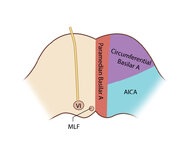 |
Figure 61: Vascular Distribution and Anatomy (Including 6th, 7th, 8th Nerves, MLF) of the Pons (Supplement) | Image | |
| 66 |
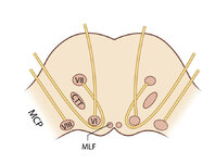 |
Figure 61: Vascular Distribution and Anatomy (Including 6th, 7th, 8th Nerves, MLF) of the Pons (Supplement) | Image | |
| 67 |
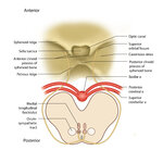 |
Figure 51: Lateral Medullary Lesion Causing Saccadic Dysmetria (Supplement) | Image | |
| 68 |
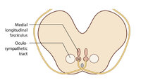 |
Figure 51: Lateral Medullary Lesion Causing Saccadic Dysmetria (Supplement) | Image | |
| 69 |
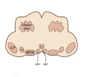 |
Figure 53: Vascular Distribution and Anatomy Relevant to the Lateral Medullary (Wallenberg) Syndrome (Supplement) | Image | |
| 70 |
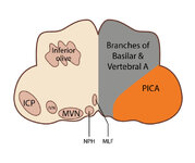 |
Figure 53: Vascular Distribution and Anatomy Relevant to the Lateral Medullary (Wallenberg) Syndrome (Supplement) | Image | |
| 71 |
 |
Figure 64: The Course of the 3rd (III) Nerve (Supplement) | Image | |
| 72 |
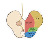 |
Figure 65: Vascular Distribution and Anatomy (Including 3rd Nerve) of the Rostral Midbrain (Supplement) | Image | |
| 73 |
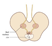 |
Figure 65: Vascular Distribution and Anatomy (Including 3rd Nerve) of the Rostral Midbrain (Supplement) | Image | |
| 74 |
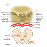 |
Figure 68: The Course of the 4th (IV) Nerve (Supplement) | Image | |
| 75 |
 |
Figure 69: Vascular Distribution and Anatomy (Including 4th Nerve) of the Caudal Midbrain (Supplement) | Image |