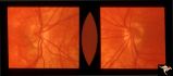
Best known for his world-renowned neuro-ophthalmology unit based at the University of California, San Francisco, William Hoyt, MD collected here more than 850 of his best images covering a wide range of disorders.
William F. Hoyt, MD, Professor Emeritus of Ophthalmology, Neurology and Neurosurgery, Department of Ophthalmology, University of California, San Francisco.
NOVEL: https://novel.utah.edu/
TO
| Title | Description | Type | ||
|---|---|---|---|---|
| 51 |
 |
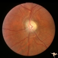
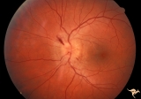
C06 Pits of the Optic Disc | Right eye. Temporal pit. 6 year old with see-saw nystagmus. Anatomy: Optic disc. Clinical: Six-year old with see-saw nystagmus. | Image |
| 52 |
 |
H32 Dysplasia with Hypoplasia (Elevated Dysplasia with Anomalous Vessels) | Left eye. 6 year old boy. Severe dysplasia. Elevated dysplasia with medullated (myelinated) nerve fibers and anomalous vessels. Son of patient in H_31 and H_10. Grandson of patient in H_11 an H_12. Anatomy: Optic disc. Pathology: Dysplasia of the optic disc. Disease/ Diagnosis: Elevated dysplasia wi... | Image |
| 53 |
 |
H23 Dysplasia with Hypoplasia (Elevated Hysplasia with Anomalous Vessels) | Elevated dysplasia with anomalous vessels. Left eye. Hypoplasia with central glial tissue remnant. Japanese girl. Same patient as H_24. Anatomy: Optic disc. Pathology: Dysplasia of the optic disc. Disease/ Diagnosis: Elevated dysplasia with hypoplasia. | Image |
| 54 |
 |
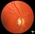
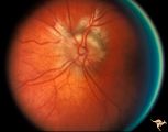
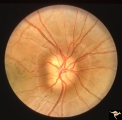
C13 Morning Glory Disc | "Morning Glory" disc with peripapillary choroidal defect extending inferiorly. Patient has transphenoidal encephalocele. Note tapering edge like an arrow pointing to patient's basal encephalocele and cleft palate. Reference: Brodsky MC, Hoyt WF, Hoyt CS, Miller NR, Lam BL. Atypical retinochoroidal ... | Image |
| 55 |
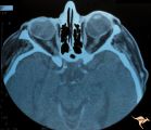 |
H41 Segmental Hypoplasia, Retinal, Tilted (Dysverted) Disc | CT scan of patient in H_40 showing marked nasal ectasia of the eyeballs. CT scan shows obliquely inserted optic nerves and marked nasal dysplasia of the eyeballs. Anatomy: Optic disc; retina. Pathology: Hypoplasia secondary to retinal lesion. Disease/ Diagnosis: Segmental optic disc hypoplasia. Imag... | Image |
| 56 |
 |
H39 Segmental Hypoplasia, Retinal, Tilted (Dysverted) Disc | Visual field of patient in H_38 showing upper temporal field depression caused by inferior nasal hypoplasia. Anatomy: Optic disc; Retina. Pathology: Hypoplasia secondary to retinal lesion. Disease/ Diagnosis: Segmental optic disc hypoplasia. Clinical: Man with bitemporal visual field defects. | Image |
| 57 |
 |
H37 Segmental Hypoplasia, Retinal, Tilted (Dysverted) Disc | Tilted (dysverted) disc in patient with high myopia. Note inferior nasal crescents with accompanying segmental hypoplasia. Man with bitemporal visual field defect. Anatomy: Optic disc, retina. Pathology: Hypoplasia secondary to retinal lesion. Disease/ Diagnosis: Segmental optic disc hypoplasia. Cli... | Image |
| 58 |
 |
H40 Segmental Hypoplasia, Retinal, Tilted (Dysverted) Disc | 60 year old woman with incidental bitemporal visual field depression. Extreme tilting of optic disc with inferior nasal segmental hypoplasia. Nasal retinal ectasia. Same patient as H_41. Anatomy: Optic disc; retina. Pathology: Hypoplasia secondary to retinal lesion. Disease/ Diagnosis: Segmental opt... | Image |
| 59 |
 |
H38 Segmental Hypoplasia; Retinal; Tilted (Dysverted) Disc | Right eye. Man with tilted (dysverted) disc with inferior nasal crescent and high myopia. Same patient as H_39. Anatomy: Optic disc; Retina. Pathology: Hypoplasia secondary to retinal lesion. Disease/ Diagnosis: Segmental optic disc hypoplasia. Clinical: Man with bitemporal visual field defects. | Image |
| 60 |
 |
Post Papilledema, Secondary Optic Atrophy | Right eye. Post papilledema with chronic gliosis. arterial narrowing. ""high-water"" marks. Man. Anatomy: Optic disc. Pathology: Post papilledema. Disease/Diagnosis: Post papilledema with optic atrophy. | Image |
| 61 |
 |
Post Papilledema, Secondary Optic Atrophy | Left eye. Post papilledema with chronic gliosis. arterial narrowing. "high-water" marks. Man. Anatomy: Optic disc. Pathology: Post papilledema. Disease/Diagnosis: Post papilledema with optic atrophy. | Image |
| 62 |
 |
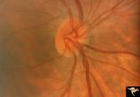
Bilateral Hemorrhagic Papilledema from Saggital Sinus Thrombosis | Left eye. 20 year old woman on oral contraceptives. Bilateral hemorrhagic Papilledema from sagittal sinus thrombosis. Anatomy: Optic disc. Pathology: Papilledema; hemorrhagic papilledema. Disease/Diagnosis: Superior saggital sinus thrombosis due to BCP use. Clinical notes: Chronic headache. | Image |
| 63 |
 |
IC102c Central Retinal Artery Occlusion with Cilioretinal Collaterals | Right eye, 1982, Central retinal artery occlusion with cilioretinal collateral occlusions due to calcific embolic occlusion behind the lamina cribrosa due to calcific valvular heart disease. Collaterals have been called "Nettleship Collaterals", recognizing the British physician who first described ... | Image |
| 64 |
 |
IC102b Central Retinal Artery Occlusion with Cilioretinal Collaterals | Right eye, 1991, Central retinal artery occlusion with cilioretinal collateral occlusions due to calcific embolic occlusion behind the lamina cribrosa due to calcific valvular heart disease. Collaterals have been called "Nettleship Collaterals", recognizing the British physician who first described ... | Image |
| 65 |
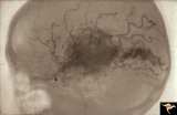 |
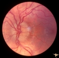
Vascular Disc Anomalies - Retinal Arteriovenous Malformations | Retinal arteriovenous malformations. Wyburn-Mason Syndrome. Angiogram showing extension of vascular malformation up the right optic nerve (arrow) through the thalamus and into the right visual cortex. References #3 and #73. Anatomy: Brain. Pathology: Arteriovenous malformation. Disease/Diagnosis: Wy... | Image |
| 66 |
 |
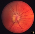
Vascular Disc Anomalies - Retinal Arteriovenous Malformations | Retinal arteriovenous malformations. Partially involved. Same patient as V_27. Anatomy: Optic disc; Brain. Pathology: Arteriovenous malformation of retina and brain. Disease/Diagnosis: Wyburn-Mason syndrome. Clinical: Blindness in the involved eye, proptosis. | Image |
| 67 |
 |
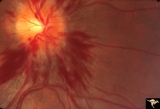
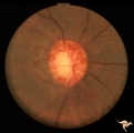
Bilateral Papilledema with Pseudotumor Cerebri | Chronic appearance of swelling in right eye. 29 year old woman. Bilateral papilledema. Anatomy: Optic disc. Pathology: Bilateral papilledema. Disease/Diagnosis: Intracranial hypertension due to treatment of growth failure with thyroid medication. Clinical: symptoms: headache, signs: bilateral papill... | Image |
| 68 |
 |
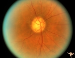
A201 Disc Swelling with Big Blind Spot Syndrome | Blind spot larger than could be explained by visible edema. Subretinal white dots probably indicate margin of blind spot. Anatomy: Optic disc; Retina. Pathology: Unknown. Disease/ Diagnosis: Big blind spot syndrome. Clinical: symptoms: photosias, blurred vision signs: Disc swelling; white spots in t... | Image |
| 69 |
 |
A202 Disc Swelling with Big Blind Spot Syndrome | Blind spot larger than could be explained by visible disc edema. Reference: Fletcher WA, Imes RK, Goodman D, Hoyt WF. Acute idiopathic blind spot enlargement. A big blind spot disc edema. Arch Ophthalmol. 1988 Jan;106(1):44-9. Anatomy: Optic disc; Retina. Pathology: Unknown. Disease/ Diagnosis: Big ... | Image |
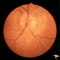
| 70 |
 |
A203 Disc Swelling with Big Blind Spot Syndrome | Slight inferior swelling in patient with grossly enlarged blind spot. 66 year old woman. Anatomy: Optic disc; Retina. Pathology: Unknown. Disease/ Diagnosis: Big blind spot syndrome. Clinical: symptoms: photopsias; blurred vision signs: disc swelling; white dots in the retina; enlarged blind spot on... | Image |
| 71 |
 |
Buried Drusen | 5 year old boy. Bilateral buried drusen. Notice the lumpy nasal disc elevation. This patient had a twin brother whose optic disc drusen were exposed. Anatomy: Optic disc. Pathology: Drusen of the optic disc. Disease/Diagnosis: Drusen of the optic disc. Clinical notes: Normally functioning eye with ... | Image |
| 72 |
 |
Buried Drusen | 5 year old boy. Bilateral buried drusen. Notice the lumpy nasal disc elevation. This patient had a twin brother whose optic disc drusen were exposed. Anatomy: Optic disc. Pathology: Drusen of the optic disc. Disease/Diagnosis: Drusen of the optic disc. Clinical notes: Normally functioning eye with ... | Image |
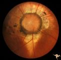
| 73 |
 |
Buried Drusen | 7 year old boy with pseudo papilledema from buried drusen. Note the lumpy contour of the disc margin. Also note the surrounding ring-like light reflex that is optically perfect and indicates absence of edema spreading onto the surrounding retina. Anatomy: Optic disc. Pathology: Drusen of the optic d... | Image |
| 74 |
 |
Buried Drusen | Left disc has a blurred lumpy margin. Retinal vessels are not obscured in the disc margin blur, therefore no edema is present. This is an example of a difficult blurred disc, the nature of which is clarified by the presence of a clear cut disk anomoly in the fellow eye. 8 year old girl. PP_15a has b... | Image |
| 75 |
 |
Buried Drusen | Buried drusen; PP_13a: Right eye. Note lumpy disc margin, especially temporally. Also note absence of optic cup. Excellent example of pseudo papilledema with buried drusen. Anatomy: Optic disc. Pathology: Drusen of the optic disc. Disease/Diagnosis: Drusen of the optic disc. Clinical notes: Patient ... | Image |