A collection of videos relating to the diagnosis and treatment of eye movement disorders. This collection includes many demonstrations of examination techniques.
Dan Gold, D.O., Associate Professor of Neurology, Ophthalmology, Neurosurgery, Otolaryngology - Head & Neck Surgery, Emergency Medicine, and Medicine, The Johns Hopkins School of Medicine.
A collection of videos relating to the diagnosis and treatment of eye movement disorders.
NOVEL: https://novel.utah.edu/
TO
| Title | Description | Type | ||
|---|---|---|---|---|
| 26 |
 |
Pons: 6th and 7th Nerve Anatomy and the Central Segmental Tract | From this cross-section of the pons, the proximity of the 6th nucleus to the 7th nerve fascicles is apparent. This is the basis of the so-called facial colliculus syndrome, where an ipsilesional horizontal gaze palsy from a nuclear 6th lesion (usually related to stroke or demyelination) can be seen ... | Image |
| 27 |
 |
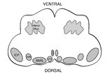
Coronal Section of the Brainstem Showing Ocular Motor Nuclei and Anatomy of the Vestibular Nucleus (with SCC Inputs) | (A) Seen here is a coronal view of the brainstem showing the locations of the ocular motor nuclei (IIIrd, IVth, VIth) as well as the nuclei of VII and VIII (vestibular and cochlear). The vestibular nucleus (VN) is divided into the inferior, lateral, medial, and superior subnuclei, and the medial ves... | Image |
| 28 |
 |
Saccadic Pathways in the Brainstem and Cerebellum & Mechanism for Saccadic Dysmetria in Wallenberg Syndrome - Normal Function of the Brainstem/Cerebellar Saccadic Pathways | The inferior cerebellar peduncle (ICP) carries climbing fibers to the dorsal vermis, and these fibers have an inhibitory influence over the Purkinje cells. These Purkinje cells normally inhibit the ipsilateral fastigial nucleus, and the fastigial nucleus projects to the contralateral inhibitory burs... | Image |
| 29 |
 |
Vestibular Neuritis with + Head Impulse Test and Unidirectional Nystagmus (Figure 1) | Vestibular neuritis is the most common cause of the acute vestibular syndrome, which is characterized by continuous vertigo and spontaneous nystagmus lasting days. It may be mimicked by central causes, including stroke, but in the hands of subspecialists, the HINTS+ (Head Impulse, Nystagmus, Test of... | Image |
| 30 |
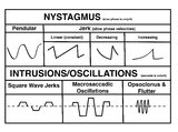 |
A Comparison of Nystagmus and Saccadic Intrusions/Oscillations | Nystagmus can be classified into pendular and jerk waveforms, where both are generated by a slow, pathologic phase. Corrective phase (the position reset mechanism) differs. In pendular nystagmus, the eyes move back and forth with about the same velocity and amplitude, similar to that of a pendulum... | Image |
| 31 |
 |
Triangle of Guillain-Mollaret | Seen here is a schematic representation of the Gullain-Mollaret triangle (Figure 1), also referred to as the dentato-olivary pathway, reflecting the 3 points of this imaginary triangle - 1) dentate nucleus, 2) red nucleus, and 3) inferior olivary nucleus. The olive sends decussating climbing fibers ... | Image |
| 32 |
 |
Medullary Structures Relevant to the Ocular Motor and Vestibular Consequences of Lateral Medullary (Wallenberg) Syndrome | This is an axial section of the medulla showing the structures that, when damaged, are responsible for the vestibular and ocular motor features of the lateral medullary or Wallenberg syndrome. The nucleus prepositus hypoglossi (NPH) and medial vestibular nucleus (MVN) complex is important for horizo... | Image |
| 33 |
 |
Saccadic Pathways in the Brainstem and Cerebellum & Mechanism for Saccadic Dysmetria in Wallenberg Syndrome - Abnormal Function of the Brainstem/Cerebellar Saccadic Pathways with a Left Wallenberg Syndrome | The end result of a lesion involving the climbing fibers within the left lateral medulla is deficient rightward saccades (contralesional hypometric saccades), and over-active leftward saccades (ipsilesional hypermetric saccades), and ipsilesional ocular lateropulsion given this baseline imbalance. M... | Image |
| 34 |
 |
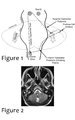
Neuro-Ophthalmic Features and Pseudo-MG Lid Signs in Miller Fisher Syndrome (Figure 1) | This is a 51-year-old woman who presented with imbalance, acute onset dizziness and diplopia that developed over three days following two weeks of upper respiratory infection and bacterial conjunctivitis. When she was initially seen as an outpatient, nystagmus was noted to the right and left, and a ... | Image |
| 35 |
 |
Subtle Torsional Pendular Nystagmus in Oculopalatal Tremor (OPT) (Figure 1) | This is a 50-year-old woman who presented with imbalance, and MRI demonstrated a right cerebellar cavernous malformation. She underwent surgery to resect the malformation, and post-operatively experienced right hemiparesis and ataxia. Six months after the surgery, balance worsened and vision became ... | Image |
| 36 |
 |
Relationship Between Semicircular Canals and Extraocular Muscles | Figure 1: When stimulated, each of the 6 angular acceleration detecting semicircular canals (3 on the right and 3 on the left) responds with a conjugate eye movement, with the vector(s) indicated below. PC=posterior canal; HC=horizontal (also known as lateral) canal; AC=anterior (also known as super... | Image |
| 37 |
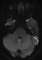 |
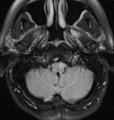
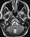
Periodic Alternating Nystagmus Due to Nodulus Stroke (Figure 1) | This is a 70-year-old woman who experienced the acute onset of vertigo and imbalance. MRI demonstrated a diffusion-weighted imaging hyperintensity involving the nodulus (with corresponding ADC hypointensity) consistent with an acute stroke. On examination several weeks after the stroke, periodic alt... | Image |
| 38 |
 |
Oculopalatal Tremor with Prominent Nystagmus, Bilateral Horizontal Gaze Palsy, and Bilateral Facial Palsies (Figure 1) | Figure 1, MRI T2 sequence demonstrating hyperintensities involving bilateral inferior olives of the medulla. This is a 50-year-old woman who experienced the acute onset of right sixth and seventh nerve palsies and left hemiparesis. Two cavernomas within the right pons (one in the region of the facia... | Image |
| 39 |
 |
The Utriculo-Ocular Motor Pathways - Physiologic and Pathologic Ocular Tilt Reaction: Physiologic Ocular Tilt Reaction (OTR) (Figure 1) | A skew deviation is a non-paralytic vertical ocular misalignment that is due to imbalance in the utriculo-ocular motor pathways. While vestibular jerk nystagmus is a consequence of static semicircular canal pathway imbalance (e.g., left-beating nystagmus due to acute right vestibular hypofunction fr... | Image |
| 40 |
 |
The Utriculo-Ocular Motor Pathways - Physiologic and Pathologic Ocular Tilt Reaction: OTR Diagram Pathologic EOMs Labelled (Figure 3) | A skew deviation is a non-paralytic vertical ocular misalignment that is due to imbalance in the utriculo-ocular motor pathways. While vestibular jerk nystagmus is a consequence of static semicircular canal pathway imbalance (e.g., left-beating nystagmus due to acute right vestibular hypofunction fr... | Image |
| 41 |
 |
The Utriculo-Ocular Motor Pathways - Physiologic and Pathologic Ocular Tilt Reaction: Pathologic OTR (Figure 2) | A skew deviation is a non-paralytic vertical ocular misalignment that is due to imbalance in the utriculo-ocular motor pathways. While vestibular jerk nystagmus is a consequence of static semicircular canal pathway imbalance (e.g., left-beating nystagmus due to acute right vestibular hypofunction fr... | Image |
| 42 |
 |
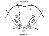
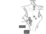
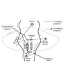
Vertical Semicircular Canal Pathways | Anterior Canal Pathway Afferents that originate in the anterior canals (AC) of the peripheral labyrinth first synapse in the ipsilateral vestibular nucleus. Three pathways exist: 1) medial longitudinal fasciculus (MLF) - right AC afferents to right medial vestibular nucleus (MVN), decussate and asc... | Image |
| 43 |
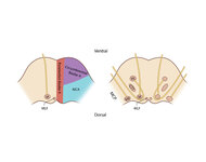 |
Figure 61: Vascular Distribution and Anatomy (Including 6th, 7th, 8th Nerves, MLF) of the Pons | In this axial section of the pons, the proximity of the 7th (VII) and 8th (VIII) fascicles can be appreciated, and a lateral inferior pontine syndrome (anterior inferior cerebellar artery, AICA territory), which could involve both of these fascicles, could cause acute prolonged vertigo accompanied b... | Image |
| 44 |
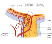 |
Figure 27: Vascular Supply of the Optic Nerve Head, Choroid and Retina | The ophthalmic artery is a branch of the internal carotid artery, which in turn, supplies the posterior ciliary (to choroid and outer retina) and central retinal (to inner retina) arteries. The central retinal artery (CRA) enters the optic nerve about 1 cm posterior to the globe, and an embolus may ... | Image |
| 45 |
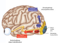 |
Figure 43: How the Brain Makes Sense of What It Sees - The Dorsal and Ventral Visual Pathways, and a 3 Tiered Approach to Vision | 1) Ventral ("what") stream - this begins with the ‘P' retinal ganglion cells à parvocellular layers of the lateral geniculate nucleus (LGN, 3-6) à V1/striate cortex (in blue) à V4/V4a (fusiform and lingual gyri) à occipitotemporal regions. 2) Dorsal ("where") stream - this begins with the ‘M... | Image |
| 46 |
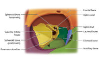 |
Figure 17: Bony Structures Relevant to the Orbit | The frontal, sphenoid, maxillary, ethmoid, and lacrimal bones make up the orbit. Structures passing through the optic canal include the optic nerve, oculosympathetic tract and ophthalmic artery. Structures passing through the superior orbital fissure include the superior ophthalmic vein, cranial ner... | Image |
| 47 |
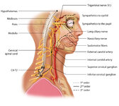 |
Figure 1: Oculosympathetic Pathway for Pupillary Dilation | The oculosympathetic tract is an uncrossed pathway that begins in the hypothalamus, with fibers descending in the brainstem (1st order, commonly affected in a lateral medullary syndrome), synapsing in the lower cervical/upper thoracic spinal cord (interomediolateral cell columns of C8-T2, also refer... | Image |
| 48 |
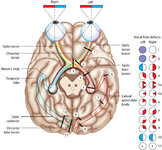 |
Figure 24: Typical Visual Field Defects Associated with Discrete Lesions Along the Visual Pathways | Specific monocular or binocular visual field defects can be highly localizing when the neuroanatomy of the visual pathways is understood. The temporal visual field corresponds to the nasal retina, while the nasal visual field corresponds to the temporal retina. 1) Left optic nerve lesion - while an ... | Image |
| 49 |
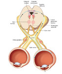 |
Figure 2: Parasympathetic Pathway for Pupillary Constriction | A bright light is shone in one eye, light enters the pupil and hyperpolarizes retinal photoreceptors which activates retinal ganglion cells. These signals propagate along the optic nerves, chiasm, optic tracts, and fibers responsible for the light reflex then synapse in the dorsal midbrain (prior to... | Image |
| 50 |
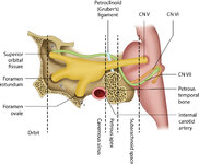 |
Figure 46: The Course of the 6th (VI) Nerve | The sixth nucleus is located dorsally, adjacent to the 4th ventricle, in the lower pons. The genu of the facial (7th) nerve wraps around the 6th nucleus, creating the facial colliculus, which bulges into the 4th ventricle. After the 6th nerve leaves the pons, it follows a vertical course along the c... | Image |