Best known for his world-renowned neuro-ophthalmology unit based at the University of California, San Francisco, William Hoyt, MD collected here more than 850 of his best images covering a wide range of disorders.
William F. Hoyt, MD, Professor Emeritus of Ophthalmology, Neurology and Neurosurgery, Department of Ophthalmology, University of California, San Francisco.
NOVEL: https://novel.utah.edu/
TO
Filters: Collection: "ehsl_novel_wfh"
| Title | Description | Type | ||
|---|---|---|---|---|
| 26 |
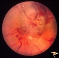 |
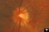
Bilateral Papilledema from Occipital Tumor | Left eye. Bilateral hemorrhagic papilledema. Occipital glioma. Woman. Anatomy: Optic disc. Pathology: Papilledema. Disease/Diagnosis: Hemorrhagic papilledema from occipital glioma. | Image |
| 27 |
 |
Bilateral Papilledema from Pseudotumor | Right eye. Chronic papilledema. Woman. Anatomy: Optic disc. Pathology: Bilateral papilledema; atrophic papilledema. Disease/Diagnosis: Pseudotumor. Clinical notes: Headache. | Image |
| 28 |
 |
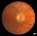
Bilateral Papilledema from Pseudotumor | Left eye. Atrophic changes in left optic disc. Chronic papilledema with involution to atrophy on the left. Woman. Anatomy: Optic disc. Pathology: Bilateal papilledema; atrophic papilledema. Disease/Diagnosis: Pseudotumor. Clinical: Headache. | Image |
| 29 |
 |
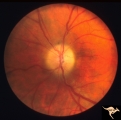
Bilateral Papilledema from Pseudotumor | Right eye. Pseudotumor syndrome. Multiple endocrine adenomas. Woman. Anatomy: Optic disc. Pathology: Bilateral papilledema. Disease/Diagnosis: Pseudotumor associated with multiple endocrine adenomas. Clinical notes: Headache; Obesity. | Image |
| 30 |
 |
Bilateral Papilledema in Pseudotumor | Left eye. Pseudotumor syndrome. Multiple endocrine adenomas. Woman. Anatomy: Optic disc. Pathology: Bilateral papilledema. Disease/Diagnosis: Pseudotumor associated with multiple endocrine adenomas. Clinical notes: Headache; Obesity. | Image |
| 31 |
 |
Bilateral Papilledema with Cyanotic Heart Disease | Bilateral Papilledema with cyanotic heart disease in a young boy. Anatomy: Optic disc. Pathology: Papilledema. Disease/Diagnosis: Pseudotumor due to cyanotic heart disease. Clinical notes: Young boy with clubbing. | Image |
| 32 |
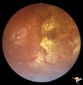 |
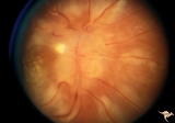
Bilateral Papilledema with Exudative Retinopathy | Right eye. Bilateral Papilledema with exudative retinopathy from vitamin A toxicity. Young boy. Near blind. Anatomy: Optic disc; Retina. Pathology: Bilateral papilledema; exudative retinopathy. Disease/Diagnosis: Hypervitaminosis A causing blindness. Clinical notes: Nearly blind; Headache. | Image |
| 33 |
 |
Bilateral Papilledema with Exudative Retinopathy | Left eye. Bilateral Papilledema with exudative retinopathy from vitamin A toxicity. Young boy. Near blind. Anatomy: Optic disc; Retina. Pathology: Bilateral papilledema; exudative retinopathy. Disease/Diagnosis: Hypervitaminosis A causing blindness. Clinical notes: Nearly blind; Headache. | Image |
| 34 |
 |
Bilateral Papilledema with Exudative Retinopathy | Left eye. Bilateral Papilledema with exudative retinopathy from vitamin A toxicity. Young boy. Near blind. Anatomy: Optic disc; Retina. Pathology: Bilateral papilledema; exudative retinopathy. Disease/Diagnosis: Hypervitaminosis A causing blindness. Clinical notes: Nearly blind; Headache. | Image |
| 35 |
 |
Bilateral Papilledema with Exudative Retinopathy | Bilateral Papilledema with exudative retinopathy from vitamin A toxicity. Young boy. Near blind. Anatomy: Optic disc; Retina. Pathology: Bilateral papilledema; exudative retinopathy. Disease/Diagnosis: Hypervitaminosis A causing blindness. Clinical notes: Nearly blind; Headache. | Image |
| 36 |
 |
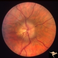
Bilateral Papilledema with Pseudotumor Cerebri | Chronic appearance of swelling in right eye. 29 year old woman. Bilateral papilledema. Anatomy: Optic disc. Pathology: Bilateral papilledema. Disease/Diagnosis: Intracranial hypertension due to treatment of growth failure with thyroid medication. Clinical: symptoms: headache, signs: bilateral papill... | Image |
| 37 |
 |
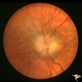
Bilateral Papilledema with Pseudotumor Cerebri | Right eye. Mild bilateral papilledema in a 7 year old boy. Cause of swelling unknown. Growth failure treated with thyroid medication. Anatomy: Optic disc. Pathology: Bilateral papilledema. Disease/Diagnosis: Intracranial hypertension due to treatment of growth failure with thyroid medicaltion. Clini... | Image |
| 38 |
 |
Bilateral Papilledema with Pseudotumor Cerebri | Left eye. Mild bilateral papilledema in a 7 year old boy. Cause of swelling unknown. Growth failure treated with thyroid medication. Anatomy: Optic disc. Pathology: Bilateral papilledema. Disease/Diagnosis: Intracranial hypertension due to treatment of growth failure with thyroid medication. Clinica... | Image |
| 39 |
 |
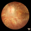
Bilateral Severe Hemorrhagic Papilledema | Right eye. Bilateral Severe Hemorrhagic Papilledema in a woman with hyperthyroidism and dural sinus occlusion. | Image |
| 40 |
 |
Bilateral Severe Hemorrhagic Papilledema | Left eye. Bilateral Severe Hemorrhagic Papilledema in a woman with hyperthyroidism and dural sinus occlusion. | Image |
| 41 |
 |
Bilateral Severe Hemorrhagic Papilledema | Right eye. 2 months later, resolving Bilateral Severe Hemorrhagic Papilledema. Same eye as P_32a | Image |
| 42 |
 |
Bilateral Severe Hemorrhagic Papilledema | Left eye. Two months later, resolving Bilateral Severe Hemorrhagic Papilledema. Same eye as P_32b | Image |
| 43 |
 |
Bilateral Severe Hemorrhagic Papilledema | Right eye. Bilateral hyperacute papilledema with rapid blindness associated with dural sinus occlusion. Both eyes were nearly blind. Young man. Anatomy: Optic disc. Pathology: Papilledema. Disease/Diagnosis: Bilateral hyperacute papilledema | Image |
| 44 |
 |
Bilateral Severe Hemorrhagic Papilledema | Left eye. Bilateral hyperacute papilledema with rapid blindess associated with dural sinus occlusion. Both eyes were nearly blind. Boy. | Image |
| 45 |
 |
Chronic Atrophic Papilledema | Right eye. Chronic Atrophic Papilledema. Obese woman (300 lbs) with large tentorial meningioma. "Pseudo Pseudotumor" Anatomy: Optic disc. Pathology: Papilledema. Disease/Diagnosis: Papilledema from large tentorial meningioma. | Image |
| 46 |
 |
Chronic Atrophic Papilledema | Left eye. Left eye blind. Chronic Atrophic Papilledema. Obese woman (300 lbs) with large tentorial meningioma. "Pseudo Pseudotumor". Anatomy: Optic disc. Pathology: Papilledema. Disease/Diagnosis: Papilledema from large tentorial meningioma. | Image |
| 47 |
 |
Chronic Papilledema due to Brain Tumor | Right eye. Chronic papilledema wth white centrally located exudates in a man with hemispheric glioma. Anatomy: Optic disc. Pathology: Papilledema. Disease/Diagnosis: Chronic papilledema. | Image |
| 48 |
 |
Chronic Papilledema due to Brain Tumor | Left eye. Chronic papilledema with white centrally located exudates in a man with hemispheric glioma. Anatomy: Optic disc. Pathology: Papilledema. Disease/Diagnosis: Chronic papilledema. | Image |
| 49 |
 |
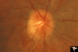
Chronic Papilledema due to Brain Tumor - Resolved | Right eye - same as P_40a - follow up after 4 months. Chronic papilledema resolved after treatment showing residual atrophy. Anatomy: Optic disc. Pathology: Papilledema. Disease/Diagnosis: Chronic papilledema. | Image |
| 50 |
 |
Chronic Papilledema due to Brain Tumor - Resolved | Left eye - same as P_40b - follow up after 4 months. Chronic papilledema resolved after treatment showing residual atrophy. | Image |