Best known for his world-renowned neuro-ophthalmology unit based at the University of California, San Francisco, William Hoyt, MD collected here more than 850 of his best images covering a wide range of disorders.
William F. Hoyt, MD, Professor Emeritus of Ophthalmology, Neurology and Neurosurgery, Department of Ophthalmology, University of California, San Francisco.
NOVEL: https://novel.utah.edu/
TO
Filters: Collection: "ehsl_novel_wfh"
| Title | Description | Type | ||
|---|---|---|---|---|
| 26 |
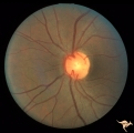 |
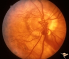
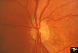
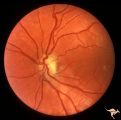
Venous Anomalies - Exit Anomalies | Disc edge veins of Kraupa. 35 year old woman. Note that the arterial branches all appear to be cilioretinal. Empty disc. Anatomy: Optic disc. Pathology: Congenital anomaly, exit anomaly. Disease/Diagnosis: Exit anomaly, edge veins. Clinical: Asymptomatic. | Image |
| 27 |
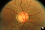 |
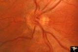
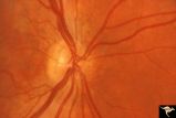
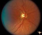
Venous Anomalies - Exit Anomalies | All venous systems drain through single vein. ""Where do they go?"" Disc edge veins of Kraupa. Anatomy: Optic disc. Pathology: Congenital anomaly, exit anomaly. Disease/Diagnosis: Exit anomaly, edge veins. Clinical: Asymptomatic. | Image |
| 28 |
 |
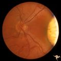
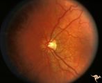
Venous Anomalies - Exit Anomalies | Anomalous exits of retinal veins at 5:00 . Anatomy: Optic disc. Pathology: Congenital anomaly, exit anomaly. Disease/Diagnosis: Exit anomaly, edge veins. Clinical: Asymptomatic. | Image |
| 29 |
 |
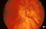
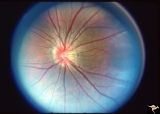
Venous Anomalies - Exit Anomalies | Choriovaginal vein. Choroid in myopic disc is raining into optic disc. Choriovaginal vein entering disc ege at 9:00. Kraupa type 2. Peripapillary atrophy in highly myopic eye. Same patient, magnified, as V_38. Anatomy: Optic disc. Pathology: Congenital anomaly of choroidal venous drainage. Disease/D... | Image |
| 30 |
 |
Venous Anomalies - Exit Anomalies | Choriovaginal vein. Choroid in myopic disc is draining into optic disc. Choriovaginal vein entering disc ege at 9:00. Kraupa type 2.Peripapillary atrophy in highly myopic eye. Same patient as V_39. Anatomy: Optic disc. Pathology: Congenital anomaly of choroidal venous drainage. Disease/Diagnosis: C... | Image |
| 31 |
 |
Venous Anomalies - Exit Anomalies | Choriovaginal vein. Inferior portion of disc at 6:00. Wide angle view of same patient as V_41. Anatomy: Optic disc. Pathology: Congenital anomaly of choroidal venous drainage. Disease/Diagnosis: Choriovaginal vein. Clinical: Asymptomatic. | Image |
| 32 |
 |
Venous Anomalies - Exit Anomalies | Choriovaginal vein. Inferior portion of disc at 5:00. Same patient as V_42. Anatomy: Optic disc. Pathology: Congenital anomaly of choroidal venous drainage. Disease/Diagnosis: Choriovaginal vein. Clinical: Asymptomatic. | Image |
| 33 |
 |
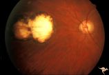
H34 Segmental Hypoplasia, Retinal-Congenital Toxo | Left eye. Temporal sector hypoplasia from congenital retinal toxoplasmosis. Note the sector shaped nerve fiber loss between 2:00 and 4:00. Same patient as H_35. Anatomy: Optic disc; retina. Pathology: Hypoplasia secondary to retinal lesion. Disease/ Diagnosis: Segmental optic disc hypoplasia. | Image |
| 34 |
 |
H35 Segmental Hypoplasia, Retinal-Congenital Toxo | Left eye. Moving out temporally to see large chorioretinal scar. Temporal sector hypoplasia from congenital retinal toxoplasmosis. Same patient as H_34. Anatomy: Optic disc; Retina. Pathology: Hypoplasia secondary to retinal lesion. Disease/ Diagnosis: Segmental optic disc hypoplasia | Image |
| 35 |
 |
H36 Segmental Hypoplasia, Retinal, Congenital Toxo | Left eye. Optic disc hypoplasia from congenital nasal retinal toxoplasma lesion. Chorioretinal scar. Anatomy: Optic disc, retina. Pathology: Hypoplasia secondary to retinal lesion. Disease/ Diagnosis: Segmental optic disc hypoplasia. | Image |
| 36 |
 |
H82 Chiasmal Hemioptic Hypoplasia | De Morsier synrome with congenital bitemporal hemianopia. Left eye. Same patient as H_81. Anatomy: Optic disc. Pathology: Chiasmal hemioptic hypoplasia. Disease/ Diagnosis: Congenital anomaly involving chiasm. | Image |
| 37 |
 |
H88 Chiasmal Hemioptic Hypoplasia | Nasal hypoplasia with temporal hemianopia from a congenital Rathke Pouch Cyst. Anatomy: Optic disc. Pathology: Chiasmal hemioptic hypoplasia. Disease/ Diagnosis: Congenital anomaly involving chiasm. | Image |
| 38 |
 |
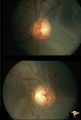
H80 Chiasmal Hemioptic Hypoplasia | Discs show striking nasal hypoplasia and band atrophy. DeMorsier synrome. Congenital bitemporal hemianopia with see-saw nystagmus. Note vertically oral shape of these hypoplastic nerves. The CT scan showed the median bar of the chiasm in this patient is totally hypoplastic. Anatomy: Optic disc. Path... | Image |
| 39 |
 |
H103 Occipital Hemianoptic Hypoplasia | Right eye. Congenital right homonymous hemianopia. Absent nerve fiber layer in right eye. Same patient as H_104. Anatomy: Optic disc. Pathology: Occipital hemianoptic hypoplasia. Disease/ Diagnosis: Congenital defect of the occipital lobe. | Image |
| 40 |
 |
H105 Occipital Hemianoptic Hypoplasia | Left congenital homonymous hemianopia. Right occipital AVM. Nasal nerve fiber layer loss in left eye. Compare with right eye. Same patient as H_106. Anatomy: Optic disc. Pathology: Occipital hemianoptic hypoplasia. DIsease/ Diagnosis: Congenital defect of the occipital lobe | Image |
| 41 |
 |
H81 Chiasmal Hemioptic Hypoplasia | De Morsier synrome with congenital bitemporal hemianopia. Right eye. Note nasal hypoplasia of the right optic disc. Same patient as H_82. Anatomy: Optic disc. Pathology: Chiasmal hemioptic hypoplasia. Disease/ Diagnosis: Congenital anomaly involving chiasm | Image |
| 42 |
 |
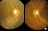
H83 Chiasmal Hemioptic Hypoplasia | De Morsier synrome with congenital bitemporal hemianopia. Note nasal hypoplasia of both optic discs. Left eye above, right eye below. Anatomy: Optic disc. Pathology: Chiasmal hemioptic hypoplasia. Disease/ Diagnosis: Congenital anomaly involving chiasm. | Image |
| 43 |
 |
H84 Chiasmal Hemioptic Hypoplasia | Congenital bitemporal hemianopia with marked bi-nasal hypoplasia. Left eye. 17 year old male. Same patient as H_85. Anatomy: Optic disc. Pathology: Chiasmal hemioptic hypoplasia. Disease/ Diagnosis: Congenital anomaly involving chiasm. | Image |
| 44 |
 |
H85 Chiasmal Hemioptic Hypoplasia | Congenital bitemporal hemianopia with marked bi-nasal hypoplasia. Right eye. 17 year old male. Same patient as H_84. Anatomy: Optic disc. Pathology: Chiasmal hemioptic hypoplasia. Disease/ Diagnosis: Congenital anomaly involving chiasm. | Image |
| 45 |
 |
H86 Chiasmal Hemioptic Hypoplasia | Congenital bitemporal hemianopia with nasal hypoplasia. 24 year old man. Same patient as H_87. Anatomy: Optic disc. Pathology: Chiasmal hemioptic hypoplasia. Disease/ Diagnosis: Congenital anomaly involving chiasm. | Image |
| 46 |
 |
H87 Chiasmal Hemioptic Hypoplasia | Congenital bitemporal hemianopia with nasal hypoplasia. 24 year old man. Same patient as H_86. Anatomy: Optic disc. Pathology: Chiasmal hemioptic hypoplasia. Disease/ Diagnosis: Congenital anomaly involving chiasm. | Image |
| 47 |
 |
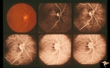
H89 Occipital Hemianoptic Hypoplasia | Diagram of homonymous hemioptic hypoplasia showing pattern of preserved nerve fibers. Homonymous hemioptic hypoplasia. Fundoscopic features in standard and red-free illumination in three patients with congenital hemiplegia. Anatomy: Optic disc. Pathology: Occipital hemianoptic hypoplasia. Disease/ D... | Image |
| 48 |
 |
PP8b Crowded Disc with Significant Nasal Disc Blurring | Congenital nasal disc blurring. Myopic eyes. Thai girl patient. One wonders about vitreal adherence to the disc. PP 8b left eye. Pair with PP 8a right eye. Anatomy: Optic disc. Pathology: Normal variation of the optic disc. Disease/ Diagnosis: Normal variation of the optic disc. Congenital blurred d... | Image |
| 49 |
 |
H101 Occipital Hemianoptic Hypoplasia | Right eye. Same patient as H_102. Anatomy: Optic disc. Pathology: Occipital hemianoptic hypoplasia. Disease/ Diagnosis: Congenital defect of the occipital lobe. | Image |
| 50 |
 |
H106 Occipital Hemianoptic Hypoplasia | Same patient as H_105. Anatomy: Optic disc. Pathology: Occipital hemianoptic hypoplasia. Disease/ Diagnosis: Congenital defect of the occipital lobe. | Image |