Best known for his world-renowned neuro-ophthalmology unit based at the University of California, San Francisco, William Hoyt, MD collected here more than 850 of his best images covering a wide range of disorders.
William F. Hoyt, MD, Professor Emeritus of Ophthalmology, Neurology and Neurosurgery, Department of Ophthalmology, University of California, San Francisco.
NOVEL: https://novel.utah.edu/
TO
Filters: Collection: "ehsl_novel_wfh"
| Title | Description | Type | ||
|---|---|---|---|---|
| 26 |
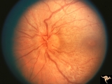 |
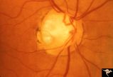
B204 Disc Swelling, Diabetic Papillopathy | Disc swelling in a diabetic. Recovered without visual loss. Left eye. Pair with B2_03. Anatomy: Optic disc. Pathology: Axoplasmic stasis due to ischemia. Disease/ Diagnosis: Diabetic papillopathy. Clinical: Visual loss with recovery. | Image |
| 27 |
 |
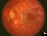
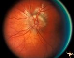
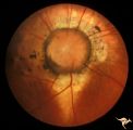
C203 Papillitis with Macular Star, Cat Scratch Disease | Proven Bartonella neuroretinitis. Left eye. October 3, 1986. Same eye as C2_04. Macular star visible on C2_04. Woman. Ocular disc edema with macular star (ODEMS). Anatomy: Optic disc; Retina. Pathology: Axoplasmic stasis due to inflammation; Exudates in Henle's layer. Disease/ Diagnosis: Bartonella ... | Image |
| 28 |
 |
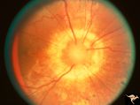
C204 Papillitis with Macular Star, Cat Scratch Disease | Proven Bartonella neuroretinitis. Left eye. October 17, 1986. Same eye as C2_03. Ocular disc edema with macular star (ODEMS). Woman. Anatomy: Optic disc; Retina. Pathology: Exudates in Henle's layer. DIsease/ Diagnosis: Neuroretinitis due to Bartonella Henslae (Cat Scratch). Clinical: Visual blurrin... | Image |
| 29 |
 |
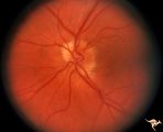
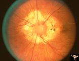
C207 Papillitis with Macular Star, Cat Scratch Disease | Proven Bartonella neuroretinitis. Anatomy: Optic disc; Retina. Pathology: Axoplasmic stasis due to inflammation. Disease/ Diagnosis: Bilateral Bartonella Henslae (Cat Scratch). Clinical: Visual blurring; Ocular disc edema with macular star (ODEMS). | Image |
| 30 |
 |
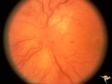
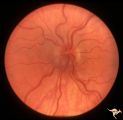
H32 Dysplasia with Hypoplasia (Elevated Dysplasia with Anomalous Vessels) | Left eye. 6 year old boy. Severe dysplasia. Elevated dysplasia with medullated (myelinated) nerve fibers and anomalous vessels. Son of patient in H_31 and H_10. Grandson of patient in H_11 an H_12. Anatomy: Optic disc. Pathology: Dysplasia of the optic disc. Disease/ Diagnosis: Elevated dysplasia wi... | Image |
| 31 |
 |
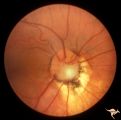
H31 Dysplasia with Hypoplasia (Elevated Hysplasia with Anomalous Vessels) | Left eye. 26 year old man. Dysplasia with hypoplasia. Father of patient in H_32. Same patient as H_10. Son of patient in H_11 an H_12. Anatomy: Optic disc. Pathology: Dysplasia of the optic disc. Disease/ Diagnosis: Elevated dysplasia with hypoplasia. | Image |
| 32 |
 |
H33 Dysplasia with Hypoplasia (Elevated Dysplasia with Anomalous Vessels) | Left eye. Elevated dysplasia, hypoplasia. Pseudo papilledema. Woman. Congenital optociliary bypass at 7:00. Anatomy: Optic disc. Pathology: Dysplasia of the optic disc. Disease/ Diagnosis: Elevated dysplasia with hypoplasia. | Image |
| 33 |
 |
H25 Dysplasia with Hypoplasia (Elevated Dysplasia with Anomalous Vessels) | Right eye. Elevated dysplasia with anomalous blood vessel pattern and peri-papillary choroidal malformation. Same patient as H_26. Anatomy: Optic disc. Pathology: Dysplasia of the optic disc. Disease/ Diagnosis: Elevated dysplasia with hypoplasia. | Image |
| 34 |
 |
H23 Dysplasia with Hypoplasia (Elevated Hysplasia with Anomalous Vessels) | Elevated dysplasia with anomalous vessels. Left eye. Hypoplasia with central glial tissue remnant. Japanese girl. Same patient as H_24. Anatomy: Optic disc. Pathology: Dysplasia of the optic disc. Disease/ Diagnosis: Elevated dysplasia with hypoplasia. | Image |
| 35 |
 |
H24 Dysplasia with Hypoplasia (Elevated Dysplasia with Anomalous Hessels) | Elevated dysplasia with anomalous vessels. Right eye. Hypoplastic with dysplasia. Japanese girl. Same patient as H_23. Anatomy: Optic disc. Pathology: Dysplasia of the optic disc. Disease/ Diagnosis: Elevated dysplasia with hypoplasia. | Image |
| 36 |
 |
H26 Dysplasia with Hypoplasia (Elevated Dysplasia with Anomalous Vessels) | Left eye. Dysplasia with grossly anomalous vascular pattern. Elevated dysplasia. Same patient as H_25. Anatomy: Optic disc. Pathology: Dysplasia of the optic disc. Disease/ Diagnosis: Elevated dysplasia with hypoplasia. | Image |
| 37 |
 |
C09 Pits of the Optic Disc | Pit with peripapillary choroidal defect. Right eye. Dwarfed boy. May not have a central retinal artery. Same patient as C_10. Anatomy: Optic disc. | Image |
| 38 |
 |
C10 Pits of the Optic Disc | Disc malformation. Abortive cavitary anomaly. Left eye. Dwarfed boy. Same patient as C_9. Anatomy: Optic disc. | Image |
| 39 |
 |
C07 Pits of the Optic Disc | Left eye. Temporal pit. Man. Anatomy: Optic disc. | Image |
| 40 |
 |
C02 Pits of the Optic Disc | Right eye. Three congenital optic pits on the temporal side. 8:00, 9:30, 10:30. Anatomy: Optic disc. | Image |
| 41 |
 |
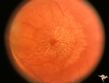
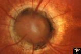
C13 Morning Glory Disc | "Morning Glory" disc with peripapillary choroidal defect extending inferiorly. Patient has transphenoidal encephalocele. Note tapering edge like an arrow pointing to patient's basal encephalocele and cleft palate. Reference: Brodsky MC, Hoyt WF, Hoyt CS, Miller NR, Lam BL. Atypical retinochoroidal ... | Image |
| 42 |
 |
C19 Morning Glory Disc | Bilateral "Morning Glory" disc. Right eye. Man. Pair with C_20. Anatomy: Optic disc. | Image |
| 43 |
 |
C20 Morning Glory Disc | Bilateral "Morning Glory" disc. Left eye. Man. Pair with C_19. Anatomy: Optic disc. | Image |
| 44 |
 |
C14 Morning Glory Disc | Isolated "Morning Glory". Left eye. Girl. Anatomy: Optic disc. | Image |
| 45 |
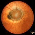 |
C11 Morning Glory Disc | "Morning Glory" disc. 11 year old girl. May not have a central retinal artery. Anatomy: Optic disc. | Image |
| 46 |
 |
C05 Pits of the Optic Disc | Right eye. Pigmented pit. Woman. Anatomy: Optic disc. | Image |
| 47 |
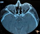 |
H41 Segmental Hypoplasia, Retinal, Tilted (Dysverted) Disc | CT scan of patient in H_40 showing marked nasal ectasia of the eyeballs. CT scan shows obliquely inserted optic nerves and marked nasal dysplasia of the eyeballs. Anatomy: Optic disc; retina. Pathology: Hypoplasia secondary to retinal lesion. Disease/ Diagnosis: Segmental optic disc hypoplasia. Imag... | Image |
| 48 |
 |
H39 Segmental Hypoplasia, Retinal, Tilted (Dysverted) Disc | Visual field of patient in H_38 showing upper temporal field depression caused by inferior nasal hypoplasia. Anatomy: Optic disc; Retina. Pathology: Hypoplasia secondary to retinal lesion. Disease/ Diagnosis: Segmental optic disc hypoplasia. Clinical: Man with bitemporal visual field defects. | Image |
| 49 |
 |
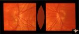
Visible Drusen | PP21a: Right eye. Drusen barely visible. Note disc margin drusen at 1:00 and 2:30.; PP21b: Left eye shows multiple exposed drusen. Girl. Anatomy: Optic disc. Pathology: Drusen of the optic disc. Disease/Diagnosis: Drusen of the optic disc. Clinical: Normally functioning eye with drusen. | Image |
| 50 |
 |
H37 Segmental Hypoplasia, Retinal, Tilted (Dysverted) Disc | Tilted (dysverted) disc in patient with high myopia. Note inferior nasal crescents with accompanying segmental hypoplasia. Man with bitemporal visual field defect. Anatomy: Optic disc, retina. Pathology: Hypoplasia secondary to retinal lesion. Disease/ Diagnosis: Segmental optic disc hypoplasia. Cli... | Image |