Collection of materials relating to neuro-ophthalmology as part of the Neuro-Ophthalmology Virtual Education Library.
NOVEL: https://novel.utah.edu/
TO
- NOVEL966
Filters: Collection: "ehsl_novel_novel"
| Title | Creator | Description | Subject | ||
|---|---|---|---|---|---|
| 26 |
 |
Optic Atrophy (PowerPoint) | William F. Hoyt, PhD | a) Evolution of optic disc pallor after optic nerve transection. Normal Right eye. Photo taken December 9, 1978. b) Injury on December 8, 1978. Evolution of optic disc pallor after optic nerve transection. Woman having rhinoplasty suffered optic nerve transection. One day after nerve transection. N... | Optic Disc Atrophy from Retrobulbar Causes (Retrograde Optic Nerve Degeneration); Severe Atrophy; Optic Atrophic |
| 27 |
 |
Temporal Atrophy (PowerPoint) | William F. Hoyt, PhD | Segmental Atrophy - Temporal pallor - Nutritional amblyopia (alcoholic). 1985. | Temporal Atrophy; Optic Disc Atrophy from Retrobulbar Causes (Retrograde Optic Nerve Degeneration); Severe Atrophy; Alcohol |
| 28 |
 |
Evolution of Optociliary Veins with Perioptic Nerve Sheath | William F. Hoyt, PhD | Series of images showing progression of disc swelling and macular degeneration. Pathology: Optociliary Vein. Disease/Diagnosis: Perioptic nerve sheath meningioma evolution. Clinical notes: Visual Loss. | Optic Disc Atrophy with Special Features; Optociliary Veins; Shunt Vessels (Meningioma) |
| 29 |
 |
Exposed Drusen (PowerPoint) | William F. Hoyt, PhD | PP25a: Left eye: Severe visual field defect. PP25b: right eye with exposed drusen and field loss: visual field defects; PP25c: right eye visual field PP25d: left eye visual field. | Pseudopapilledema; Exposed Drusen |
| 30 |
 |
Curtain Sign (Enhanced Ptosis) - Associated Image 2 | Bashaer Aldhahwani, MD; Hong Jiang, MD, PhD | This is a 78-year-old male patient who presented with diplopia, right eyelid ptosis, and ophthalmoplegia. He had severe ptosis OD and pseudo-proptosis (lid retraction) OS at baseline, but when the right eyelid was manually elevated, there was marked enhanced ptosis of the left eyelid (Video). He was... | Myasthenia GravIs; Clinical Signs |
| 31 |
 |
Myelinated Retinal Nerve Fiber Layer | Bashaer Aldhahwani, MD; Hong Jiang, MD, PhD | A 78 YOF with no visual symptoms has an incidental finding of yellow-white well-demarcated patches with ragged borders at the peripapillary area of her left eye (see the fundus photo). | Myelinated Retinal Nerve Fiber Layer |
| 32 |
 |
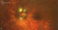
Central Retinal Artery Occlusion | Natasha Nayak, MD; Rudrani Banik, MD | Power point of case presentation of acute central retinal artery occlusion (CRAO) treated with tPA. Risk factors for stroke and results of EAGLE study reviewed. Imaging: Number of Figures and legend for each: 12 Slide 3: Figure 1: Table 1: Exam Findings Slide 3: Figure 2: Table 2: Exam Findings Cont... | Central Retinal Artery Occlusion; Stroke; Tissue Plasminogen Activator; EAGLE Study |
| 33 |
 |
Carotid Cavernous Fistula | Adam Botwinick, MD; Rudrani Banik, MD | Power point of case presentation of 66-year-old female with chronic red eye OU x 2 months, misdiagnosed as conjunctivitis. Exam showed dilated, tortuous episcleral vessels OU with proptosis OU and elevated intraocular pressure. MRI showed suspicion of carotid cavernous fistula (CCF), confirmed by ... | Carotid Cavernous Fistula; Dural CCF; Chemosis; Corkscrew Vessels; Proptosis; Embolization; Neurointerventional Radiology |
| 34 |
 |
Bitemporal Hemianopia | Julia Mathew Padiyedathu, MD; Rudrani Banik, MD | Power point of case presentation of patient with painless progressive vision loss, optic nerve cupping with pallor and history of significant alcohol and tobacco use. Patient initially diagnosed at outside institution with normal tension glaucoma and toxic optic neuropathy. Exam suggests bitempora... | Ditemporal Visual Field Defect; Toxic Optic Neuropathy; Pituitary Adenoma; Compressive Optic Neuropathy |
| 35 |
 |
Pupillary reflex and the APD | Wade Crow, MD | Illustrations describing pupillary reflex. | Pupillary Reflex, APD |
| 36 |
 |
Optic Nerve Hypoplasia (ONH) - Double Ring Sign | Bashaer Aldhahwani, MD; Joshua Pasol, MD | Optic nerve hypoplasia (ONH) is characterized by a decreased number of optic nerve axons. It can present unilaterally or bilaterally, Isolated or associated with midline cerebral structural defects, such as septum pellucidum absence, agenesis of corpus callosum, cerebral hemisphere abnormalities, or... | Optic Nerve Hypoplasia (ONH) |
| 37 |
 |
Supranuclear and Infranuclear Motility Disorder | Brittany Lin, MD; Rudrani Banik, MD | Power point of case presentation of patient with supranuclear left gaze preference from frontotemporal CVA (overcome by Doll's head), as well as right sixth nerve palsy with incomitant esotropia from pontine CVA. | Supranuclear Gaze Palsy; Sixth Nerve Palsy; Esotropia; Gaze Preference; Stroke |
| 38 |
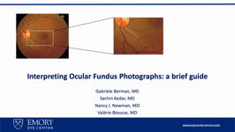 |
Interpreting Ocular Fundus Photographs: A Brief Guide | Gabriele Berman; Sachin Kedar; Nancy J. Newman; Valerie Biousse | This is a brief guide to the interpretation of the ocular fundus photograph. In this presentation we will describe the structures that comprise the normal ocular fundus followed the abnormalities that can be detected on fundus photographs. By the end of the presentation, learners should be able to d... | Fundus Photograph; Glaucoma; Papilledema; Retinal Detachment |
| 39 |
 |
Dry Eye Syndrome (Spanish) | NANOS | People with abnormalities of the tear film are diagnosed with "dry eyes", but some patients with "dry eyes" may not feel that their eyes are "dry". Itching, burning, a scratchy sensation, a sensation that there is sand or grit in the eye, or intermittent blurring of the vision can all be symptoms of... | Dry Eye Syndrome; Patient Brochure |
| 40 |
 |
Brain MRI in Multiple Sclerosis (Guest Lecture) | Anne G. Osborn, MD | The patient is a 25 year old woman who was in excellent health until 4 days prior to admission when she noted blurred vision and horizontal double vision on lateral gaze to right and left. Past History: Negative for strabismus as a child. No previous episodes of transient neurological symptoms. Fami... | Bilateral Internuclear Ophthalmoplegia; Abducting Nystagmus; Normal Convergence; Gaze Evoked Upbeat Nystagmus; Gaze Evoked Downbeat Nystagmus; Saccadic Dysmetria; Multiple Sclerosis; Horizontal Saccadic Dysmetria |
| 41 |
 |
Pendular Vertical Oscillations | Shirley H. Wray, MD, PhD, FRCP | See also: http://content.lib.utah.edu/cdm/ref/collection/ehsl-shw/id/68, http://content.lib.utah.edu/cdm/ref/collection/ehsl-shw/id/247, http://content.lib.utah.edu/cdm/ref/collection/ehsl-shw/id/111, http://content.lib.utah.edu/cdm/ref/collection/ehsl-shw/id/312, http://content.lib.utah.edu/cdm/ref... | Palatal Tremor (Myoclonus); Pendular Vertical Oscillations; Unilateral Horizontal Gaze Palsy; Facial Palsy; Pontine Infarct; Degenerative Hypertrophy of the Inferior Olivary Nucleus; Lesion in the Guillain-Mollaret Triangle; Oculopalatal Myoclonus; Oculopalatal Tremor Lid Nystagmus; Bilater... |
| 42 |
 |
Dry Eye Syndrome | NANOS | People with abnormalities of the tear film are diagnosed with "dry eyes", but some patients with "dry eyes" may not feel that their eyes are "dry". Itching, burning, a scratchy sensation, a sensation that there is sand or grit in the eye, or intermittent blurring of the vision can all be symptoms of... | Dry Eye Syndrome; Patient Brochure |
| 43 |
 |
Paraneoplastic Upbeat Nystagmus | Shirley H. Wray, MD, PhD, FRCP | This case was presented to the Clinical Eye Movement Society at the American Neurological Association Meeting in October 2009. The patient is a 65 year old woman who was in good health until seven weeks prior to admission. On June 22/09 on the return flight from her daughter's wedding in Oregon she ... | Upbeat Nystagmus; Lid Nystagmus; Square Wave Jerks (Saccadic Oscillations); Saccadic Dysmetria; Saccadic Pursuit; Paraneoplastic Syndrome; Paraneoplastic Upbeat Nystagmus; Pancreatic Endocrine Carcinoma; Anti-Hu - Associated Paraneoplastic Encephalitis |
| 44 |
 |
Oculomasticatory Myorhythmia | Shirley H. Wray, MD, PhD, FRCP | This case, previously reported in 1986, is published courtesy of John Selhorst, M.D., Saint Louis University School of Medicine, St. Louis, MO. (4) The patient is a 46 year old man who, over a period of six months, lost the ability to read and complained of excessive somnolence, occasional urinary i... | Somnolence; Supranuclear Paralysis of Up and Downgaze; Pendular Vergence Oscillations; Oculomasticatory Myorhythmia; Tropheryma Whippelii - Infection; CNS Whipple's Disease; Supranuclear Paralysis of Up and Downgaze Infection-Whipple's Disease; Oculomasticatory Myorhythmia (Whipple's) |
| 45 |
 |
One and a Half Syndrome | Shirley H. Wray, MD, PhD, FRCP | This 44 year old woman presented in 1973 with an acute attack of optic neuritis in the right eye that fully recovered after a course of ACTH therapy. In 1991, 18 years later, she developed unsteadiness of gait, "walking like a chicken", stiff legs that jerked spontaneously in bed at night, and numbn... | Unilateral Internuclear Ophthalmoplegia; Unilateral Horizontal Gaze Palsy; Upbeat Nystagmus on Upgaze; Convergence Normal; Fisher's One-and-a-Half Syndrome; Uhthoff's Symptom; Multiple Sclerosis; Unilateral Horizontal Gaze Palsy Demyelination; Gaze Evoked Upbeat Nystagmus; Abducting Nystagmus |
| 46 |
 |
Pendular Oscillations | Shirley H. Wray, MD, PhD, FRCP | This 37 year old woman has had progressive multiple sclerosis (MS) affecting the cerebellum and brainstem for 6 years. Neurological examination: Titubation Dysarthria Incoordination of the extremities Ataxic gait Spastic paraparesis with generalized hyperreflexia and extensor plantar responses. Neur... | Pendular Horizontal Oscillations; Left Beating Nystagmus; Obtrusive Saccades; Bilateral Gaze Evoked Nystagmus; Saccadic Dysmetria; Oscillopsia; Titubation; Ataxia; Multiple Sclerosis; Primary Position Horizontal Nystagmus; Gaze Evoked Horizontal Nystagmus; Horizontal Saccadic Dysmetria |
| 47 |
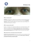 |
Anisocoria | NANOS | Anisocoria is a medical term for unequal pupil size. Normally our pupils are relatively the same size. While small differences in pupil size are normal and can even come and go ( physiologic anisocoria ), constant and significant differences in pupil sizes may be a sign of damage to the nerves that ... | Anisocoria; Patient Brochure |
| 48 |
 |
Homonymous Hemianopia | NANOS | This refers to an absence of vision towards one side of the visual world in each eye. The damage that caused this problem is in the brain and not in the eyes. Updated April 2020. | Homonymous Hemianopia; Patient Brochure |
| 49 |
 |
Optic Disc Drusen | NANOS | Optic disc drusen are abnormal deposits of protein-like material in the optic disc - the front part of the optic nerve. Updated April 2020. | Optic Disc Drusen; Patient Brochure |
| 50 |
 |
Lateropulsion | Shirley H. Wray, MD, PhD, FRCP | This 60 year old patient has Wallenberg's syndrome due to infarction of the left dorsolateral medulla. Wallenberg's syndrome is the best recognized syndrome involving the vestibular nuclei and adjacent structures. Unilateral infarcts affecting the vestibular nuclei may produce an oculomotor imbalanc... | Deviation of the Eyes Under Closed Lids; Lateropulsion; Dorsolateral Medullary Infarction; Medulla Infarct |