A collection of videos relating to the diagnosis and treatment of eye movement disorders. This collection includes many demonstrations of examination techniques.
Dan Gold, D.O., Associate Professor of Neurology, Ophthalmology, Neurosurgery, Otolaryngology - Head & Neck Surgery, Emergency Medicine, and Medicine, The Johns Hopkins School of Medicine.
A collection of videos relating to the diagnosis and treatment of eye movement disorders.
NOVEL: https://novel.utah.edu/
TO
| Title | Description | Type | ||
|---|---|---|---|---|
| 26 |
 |
Apraclonidine Testing in Horner's syndrome | This patient experienced relatively abrupt ptosis and was seen and diagnosed with a Horner's syndrome within a few days of the onset. There were no other exam findings and history did not offer clues as to the etiology. Neuroimaging of the oculosympathetic tract was unrevealing. Apraclonidine testin... | Image/MovingImage |
| 27 |
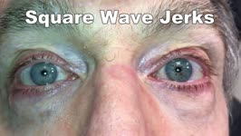 |
Saccadic Intrusions (Square Wave Jerks, SWJ) | 𝗢𝗿𝗶𝗴𝗶𝗻𝗮𝗹 𝗗𝗲𝘀𝗰𝗿𝗶𝗽𝘁𝗶𝗼𝗻: Seen here are SWJ, which is the most common example of a saccadic intrusion. Here the patient is fixating on the camera, and all of the sudden a saccade takes the eyes off the fixation target, there's a brief intersaccadi... | Image/MovingImage |
| 28 |
 |
Head Movement Independent ('Sitting') Oscillopsia - A Common Symptom of Nystagmus and Saccadic Intrusions/Oscillations | 𝗢𝗿𝗶𝗴𝗶𝗻𝗮𝗹 𝗗𝗲𝘀𝗰𝗿𝗶𝗽𝘁𝗶𝗼𝗻: This video is an example of what a patient with spontaneous nystagmus or saccadic intrusions/oscillations experiences visually during the abnormal eye movements - i.e., oscillopsia (illusion of movement of the stationary ... | Image/MovingImage |
| 29 |
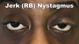 |
Jerk Nystagmus | 𝗢𝗿𝗶𝗴𝗶𝗻𝗮𝗹 𝗗𝗲𝘀𝗰𝗿𝗶𝗽𝘁𝗶𝗼𝗻: This is an example of jerk nystagmus due to a central vestibular lesion. The slow phase is the pathologic phase (to the left) which initiates the movement, and is followed by a fast position reset mechanism (to the right)... | Image/MovingImage |
| 30 |
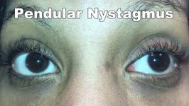 |
Pendular Nystagmus | 𝗢𝗿𝗶𝗴𝗶𝗻𝗮𝗹 𝗗𝗲𝘀𝗰𝗿𝗶𝗽𝘁𝗶𝗼𝗻: This is an example of pendular nystagmus, where like jerk nystagmus, the slow phase initiates the movement. However, unlike jerk nystagmus, there is no fast phase, but rather back to back slow phases resembling a pendulum... | Image/MovingImage |
| 31 |
 |
Horizontal Gaze Palsy, Facial Nerve Palsy, and Nystagmus Due to Dorsal Pontine Ischemia | 𝗢𝗿𝗶𝗴𝗶𝗻𝗮𝗹 𝗗𝗲𝘀𝗰𝗿𝗶𝗽𝘁𝗶𝗼𝗻: Presented here are two patients with horizontal gaze and facial palsies due to stroke. The first patient is a 60-year-old man who presented with double vision and hemiparesis due to a right dorsal pontine ischemic stroke.... | Image/MovingImage |
| 32 |
 |
Downbeat Nystagmus and Convergence Spasm | This is a 60-yo-woman with vertical oscillopsia related to her downbeat nystagmus, and diplopia related to an intermittent esotropia. When the esotropia was present, with versions there were bilateral abduction deficits. With ductions and the vestibulo-ocular reflex, it was apparent that the range o... | Image/MovingImage |
| 33 |
 |
Idiopathic Downbeat Nystagmus, Decreasing with Convergence | This is a 25-yo-woman who experienced vertically oscillopsia for 1 year, and was found to have downbeat nystagmus. Interestingly, there were no other cerebellar ocular motor signs - e.g., normal saccades, smooth pursuit, VOR suppression, and no gaze-evoked nystagmus, although her (pure) downbeat was... | Image/MovingImage |
| 34 |
 |
Cerebellar Degeneration with Downbeat Nystagmus Provoked by Convergence | Description: This is a 70-yo-woman with a progressive gait disorder, diagnosed with cerebellar ataxia. She displayed typical cerebellar ocular motor signs including gaze-evoked nystagmus, choppy pursuit and VOR suppression, and there was very subtle spontaneous downbeat nystagmus, best appreciated w... | Image/MovingImage |
| 35 |
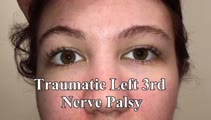 |
Traumatic 3rd Nerve Palsy with Aberrant Regeneration | 𝗢𝗿𝗶𝗴𝗶𝗻𝗮𝗹 𝗗𝗲𝘀𝗰𝗿𝗶𝗽𝘁𝗶𝗼𝗻: This is a 20-yo-woman who experienced severe head trauma and diplopia upon awakening from a coma several weeks after the injury. She had a partial left 3rd nerve palsy (adduction spared), and when she looked to the right ... | Image/MovingImage |
| 36 |
 |
Aberrant Regeneration of the 3rd Nerve | Aberrant regeneration in two patients: 1) a young woman with a right cavernous sinus meningioma with subsequent development of aberrant regeneration demonstrated by eyelid elevation OD in attempted downgaze (i.e., some fibers that were supposed to innervate the right IR were misrouted to the right l... | Image/MovingImage |
| 37 |
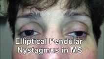 |
Elliptical Pendular Nystagmus in MS | 𝗢𝗿𝗶𝗴𝗶𝗻𝗮𝗹 𝗗𝗲𝘀𝗰𝗿𝗶𝗽𝘁𝗶𝗼𝗻: This is a 40-yo-woman with MS and bilateral optic nerve disease who presented with a year's long history of oscillopsia, which was related to elliptical pendular nystagmus. The appearance of elliptical nystagmus is the re... | Image/MovingImage |
| 38 |
 |
Idiopathic Downbeat Nystagmus Exacerbated with Positional Maneuvers | 𝗢𝗿𝗶𝗴𝗶𝗻𝗮𝗹 𝗗𝗲𝘀𝗰𝗿𝗶𝗽𝘁𝗶𝗼𝗻: This is a 45-yo-woman with vertical oscillopsia for 6+ months, found to have downbeat nystagmus on examination. She mainly complained of dizziness and oscillopsia when laying down. She was found to have a significant exac... | Image/MovingImage |
| 39 |
 |
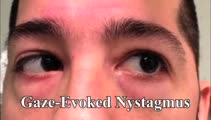
Cerebellar Eye Signs in SCA8 | 𝗢𝗿𝗶𝗴𝗶𝗻𝗮𝗹 𝗗𝗲𝘀𝗰𝗿𝗶𝗽𝘁𝗶𝗼𝗻: This is a 30-yo-man with a diagnosis of SCA 8 who had appendicular and gait ataxia in addition to choppy smooth pursuit and VORS, downbeat nystagmus, saccadic hypermetria, and gaze-evoked nystagmus with rebound nystagmus.... | Image/MovingImage |
| 40 |
 |
Vertical-Torsional Pendular Nystagmus and Convergence Spasm Due to Anti-MaTa Encephalitis | This is a 50-yo-woman with debilitating oscillopsia due to a high frequency (6 Hz) vertical-torsional pendular (quantitative eye movement recordings were performed) nystagmus. She also had intermittent double vision due to (organic) convergence spasm. Her nystagmus and spasm were thought to be relat... | Image/MovingImage |
| 41 |
 |
Gaze-evoked and Rebound Nystagmus in a Cerebellar Syndrome | 𝗢𝗿𝗶𝗴𝗶𝗻𝗮𝗹 𝗗𝗲𝘀𝗰𝗿𝗶𝗽𝘁𝗶𝗼𝗻: 30-yo-man with the subacute onset of a cerebellar syndrome. After extensive evaluation and progression, it was thought that this represented an autoimmune process and there was some improvement with immunosuppression. He ... | Image/MovingImage |
| 42 |
 |
Anterior Canal - BPPV: Deep Head Hanging | 𝗢𝗿𝗶𝗴𝗶𝗻𝗮𝗹 𝗗𝗲𝘀𝗰𝗿𝗶𝗽𝘁𝗶𝗼𝗻: Regardless or whether it is thought that the patient has right or left anterior canal (AC) involvement, the deep head hanging maneuver is performed in the same way. • First the patient is placed in the long-sitting posi... | Image/MovingImage |
| 43 |
 |
Enhanced Ptosis in Myasthenia Gravis | This is a 20-yo-woman who presented with generalized weakness, ptosis and ophthalmoplegia. She had severe ptosis OU at baseline, but when one eyelid was manually elevated, there was marked enhanced ptosis of the opposite eyelid. This was in accordance with Hering's law of equal innervation of the le... | Image/MovingImage |
| 44 |
 |
Miller Fisher Syndrome - Ophthalmoplegia and Hyperreflexia | 𝗢𝗿𝗶𝗴𝗶𝗻𝗮𝗹 𝗗𝗲𝘀𝗰𝗿𝗶𝗽𝘁𝗶𝗼𝗻: This is a 45-yo-woman who presented with mild imbalance and diplopia. There had been a preceding viral illness several weeks prior. Examination demonstrated horizontal gaze paresis (sparing unilateral adduction), mild gai... | Image/MovingImage |
| 45 |
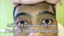 |
Miller Fisher Syndrome - Ophthalmoplegia, Ptosis and Ataxia | 𝗢𝗿𝗶𝗴𝗶𝗻𝗮𝗹 𝗗𝗲𝘀𝗰𝗿𝗶𝗽𝘁𝗶𝗼𝗻: This is a young man who presented with ptosis, difficulty moving the eyes and gait imbalance several weeks after a GI illness. Miller Fisher syndrome was diagnosed, IVIG therapy was initiated, and anti-Gq1b antibodies cam... | Image/MovingImage |
| 46 |
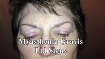 |
Typical Lid Signs (Cogan's Lid Twitch, Lid Hopping, Enhanced Ptosis) in Myasthenia Gravis | 𝗢𝗿𝗶𝗴𝗶𝗻𝗮𝗹 𝗗𝗲𝘀𝗰𝗿𝗶𝗽𝘁𝗶𝗼𝗻: This is a 60-yo-woman with MG who displays typical eyelid signs including Cogan's lid twitch, lid hopping (appreciated during horizontal smooth pursuit in this patient), and enhanced ptosis in accordance with Hering's law... | Image/MovingImage |
| 47 |
 |
Prolonged Lid Twitch in Myasthenia Gravis | This 50-yo-woman with ocular MG demonstrated a spontaneous and particularly prolonged eyelid twitch. | Image/MovingImage |
| 48 |
 |
Wall-eyed Bilateral INO in Caudal Midbrain Lesion | This is a 30-yo-woman with the relatively acute onset of diplopia. There was a large angle exotropia, very subtle lag of the adducting saccades OD>OS, suggestive of bilateral INOs. This was best seen with rapid horizontal saccades, and a lesion involving bilateral MLFs in the caudal midbrain was dem... | Image/MovingImage |
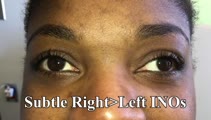
| 49 |
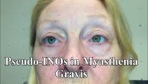 |
Pseudo-INOs in Myasthenia Gravis | This is a 55-yo-woman with an intermittent exotropia who had normal adduction OU, but clear lag of adducting saccades OD>OS with rapid horizontal saccades. This was much more apparent after repeat testing (ie, it was fatigable), and she wound up having ocular MG. | Image/MovingImage |
| 50 |
 |
One-and-a-Half Syndrome, Facial Palsy, and Nystagmus Due to Dorsal Pontine Demyelination | This is a 16-yo-girl with oscillopsia and double vision. Exam showed inability to look to the left with either eye due to left nuclear 6th. There was also a left INO (horizontal gaze palsy + INO = one-and-a-half syndrome) from left MLF involvement and left lower motor neuron facial palsy due to fasc... | Image/MovingImage |