A collection of videos relating to the diagnosis and treatment of eye movement disorders. This collection includes many demonstrations of examination techniques.
Dan Gold, D.O., Associate Professor of Neurology, Ophthalmology, Neurosurgery, Otolaryngology - Head & Neck Surgery, Emergency Medicine, and Medicine, The Johns Hopkins School of Medicine.
A collection of videos relating to the diagnosis and treatment of eye movement disorders.
NOVEL: https://novel.utah.edu/
TO
Filters: Collection: "ehsl_novel_gold"
| Title | Description | Type | ||
|---|---|---|---|---|
| 26 |
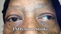 |
INOs in Stroke | 𝗢𝗿𝗶𝗴𝗶𝗻𝗮𝗹 𝗗𝗲𝘀𝗰𝗿𝗶𝗽𝘁𝗶𝗼𝗻: This video shows 3 patients with vascular risk factors who suffered strokes of the MLF resulting in unilateral INO in each case. In the second case, INO was diagnosed status post cardiac catherization and MRI was found to... | Image/MovingImage |
| 27 |
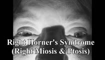 |
Horner's Syndrome with Anhidrosis | 𝗢𝗿𝗶𝗴𝗶𝗻𝗮𝗹 𝗗𝗲𝘀𝗰𝗿𝗶𝗽𝘁𝗶𝗼𝗻: This is a patient with the onset of ptosis OD years prior, with clear evidence of a Horner's syndrome. Imaging of the oculosympathetic tract was unrevealing. The patient also mentioned that with exercise, the left side of... | Image/MovingImage |
| 28 |
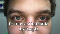 |
Eye Signs in Infantile Esotropia - Latent Nystagmus and Inferior Oblique Overaction | 𝗢𝗿𝗶𝗴𝗶𝗻𝗮𝗹 𝗗𝗲𝘀𝗰𝗿𝗶𝗽𝘁𝗶𝗼𝗻: This is a 25-yo-man with a history of amblyopia and intermittent eye crossing. On exam, he had a comitant 25 prism diopter esotropia, and other features of infantile (or congenital) esotropia including: latent nystagmus (... | Image/MovingImage |
| 29 |
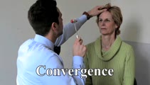 |
Convergence | Can bring out or change the direction of vertical nystagmus in Wernicke's, or cerebellar disease; may be impaired in Parkinson's disease, head trauma, elderly patients; may overcome an adduction deficit with an INO. Instructional ocular motor examination procedures. | Image/MovingImage |
| 30 |
 |
Apraclonidine Testing in Horner's syndrome | This patient experienced relatively abrupt ptosis and was seen and diagnosed with a Horner's syndrome within a few days of the onset. There were no other exam findings and history did not offer clues as to the etiology. Neuroimaging of the oculosympathetic tract was unrevealing. Apraclonidine testin... | Image/MovingImage |
| 31 |
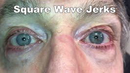 |
Saccadic Intrusions (Square Wave Jerks, SWJ) | 𝗢𝗿𝗶𝗴𝗶𝗻𝗮𝗹 𝗗𝗲𝘀𝗰𝗿𝗶𝗽𝘁𝗶𝗼𝗻: Seen here are SWJ, which is the most common example of a saccadic intrusion. Here the patient is fixating on the camera, and all of the sudden a saccade takes the eyes off the fixation target, there's a brief intersaccadi... | Image/MovingImage |
| 32 |
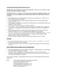 |
An Optokinetic Stimulation Home Exercise Program | 𝗢𝗿𝗶𝗴𝗶𝗻𝗮𝗹 𝗗𝗲𝘀𝗰𝗿𝗶𝗽𝘁𝗶𝗼𝗻: A plainly written program of optokinetic exercise intended for patient use at home. 𝗡𝗲𝘂𝗿𝗼-𝗼𝗽𝗵𝘁𝗵𝗮𝗹𝗺𝗼𝗹𝗼𝗴𝘆 𝗮𝗻𝗱 𝗡𝗲𝘂𝗿𝗼-𝗼𝘁𝗼𝗹𝗼𝗴𝘆 ... | Text |
| 33 |
 |
Expanded Acute Onset Persistent Vision Loss Differential | Text | |
| 34 |
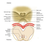 |
Figure 51: Lateral Medullary Lesion Causing Saccadic Dysmetria (Supplement) | Image | |
| 35 |
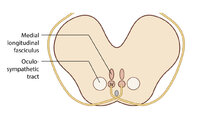 |
Figure 51: Lateral Medullary Lesion Causing Saccadic Dysmetria (Supplement) | Image | |
| 36 |
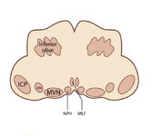 |
Figure 53: Vascular Distribution and Anatomy Relevant to the Lateral Medullary (Wallenberg) Syndrome (Supplement) | Image | |
| 37 |
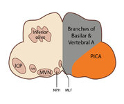 |
Figure 53: Vascular Distribution and Anatomy Relevant to the Lateral Medullary (Wallenberg) Syndrome (Supplement) | Image | |
| 38 |
 |
Figure 64: The Course of the 3rd (III) Nerve (Supplement) | Image | |
| 39 |
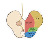 |
Figure 65: Vascular Distribution and Anatomy (Including 3rd Nerve) of the Rostral Midbrain (Supplement) | Image | |
| 40 |
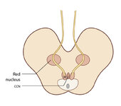 |
Figure 65: Vascular Distribution and Anatomy (Including 3rd Nerve) of the Rostral Midbrain (Supplement) | Image | |
| 41 |
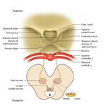 |
Figure 68: The Course of the 4th (IV) Nerve (Supplement) | Image | |
| 42 |
 |
Figure 69: Vascular Distribution and Anatomy (Including 4th Nerve) of the Caudal Midbrain (Supplement) | Image | |
| 43 |
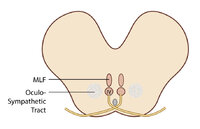 |
Figure 69: Vascular Distribution and Anatomy (Including 4th Nerve) of the Caudal Midbrain (Supplement) | Image | |
| 44 |
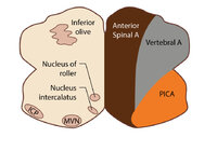 |
Figure 80: Vascular Distribution and Anatomy Relevant to the Medial Medullary Syndrome (Supplement) | Image | |
| 45 |
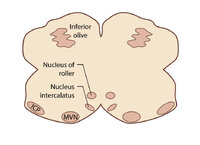 |
Figure 80: Vascular Distribution and Anatomy Relevant to the Medial Medullary Syndrome (Supplement) | Image | |
| 46 |
 |
Head Movement Independent ('Sitting') Oscillopsia - A Common Symptom of Nystagmus and Saccadic Intrusions/Oscillations | 𝗢𝗿𝗶𝗴𝗶𝗻𝗮𝗹 𝗗𝗲𝘀𝗰𝗿𝗶𝗽𝘁𝗶𝗼𝗻: This video is an example of what a patient with spontaneous nystagmus or saccadic intrusions/oscillations experiences visually during the abnormal eye movements - i.e., oscillopsia (illusion of movement of the stationary ... | Image/MovingImage |
| 47 |
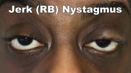 |
Jerk Nystagmus | 𝗢𝗿𝗶𝗴𝗶𝗻𝗮𝗹 𝗗𝗲𝘀𝗰𝗿𝗶𝗽𝘁𝗶𝗼𝗻: This is an example of jerk nystagmus due to a central vestibular lesion. The slow phase is the pathologic phase (to the left) which initiates the movement, and is followed by a fast position reset mechanism (to the right)... | Image/MovingImage |
| 48 |
 |
Localization of Ophthalmoplegia | 𝗢𝗿𝗶𝗴𝗶𝗻𝗮𝗹 𝗗𝗲𝘀𝗰𝗿𝗶𝗽𝘁𝗶𝗼𝗻: A table describing the localization of ophthalmoplegia. | Text |
| 49 |
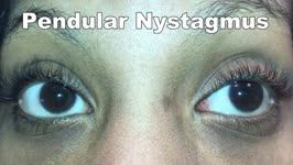 |
Pendular Nystagmus | 𝗢𝗿𝗶𝗴𝗶𝗻𝗮𝗹 𝗗𝗲𝘀𝗰𝗿𝗶𝗽𝘁𝗶𝗼𝗻: This is an example of pendular nystagmus, where like jerk nystagmus, the slow phase initiates the movement. However, unlike jerk nystagmus, there is no fast phase, but rather back to back slow phases resembling a pendulum... | Image/MovingImage |
| 50 |
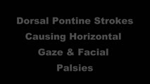 |
Horizontal Gaze Palsy, Facial Nerve Palsy, and Nystagmus Due to Dorsal Pontine Ischemia | 𝗢𝗿𝗶𝗴𝗶𝗻𝗮𝗹 𝗗𝗲𝘀𝗰𝗿𝗶𝗽𝘁𝗶𝗼𝗻: Presented here are two patients with horizontal gaze and facial palsies due to stroke. The first patient is a 60-year-old man who presented with double vision and hemiparesis due to a right dorsal pontine ischemic stroke.... | Image/MovingImage |