Best known for his world-renowned neuro-ophthalmology unit based at the University of California, San Francisco, William Hoyt, MD collected here more than 850 of his best images covering a wide range of disorders.
William F. Hoyt, MD, Professor Emeritus of Ophthalmology, Neurology and Neurosurgery, Department of Ophthalmology, University of California, San Francisco.
NOVEL: https://novel.utah.edu/
TO
| Title | Description | Type | ||
|---|---|---|---|---|
| 26 |
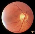 |
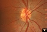
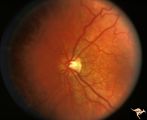
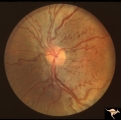
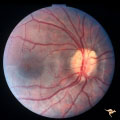
C38 Anomalous Pale Disc | Megalopapilla in -8 myopic eye. Right eye. Anatomy: Optic disc. Clinical: High myope. | Image |
| 27 |
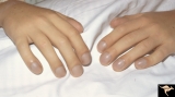 |
Cyanotic Heart Disease with Clubbing of Fingernails | Note the cyanotic nail beds and clubbing. Anatomy: Optic disc. Pathology: Papilledema. Disease/Diagnosis: Pseudotumor due to cyanotic heart disease. Clinical: Young boy with clubbing. | Image |
| 28 |
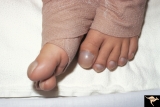 |
Cyanotic Heart Disease with Clubbing of Toes | Bilateral Papilledema with cyanotic heart disease. Anatomy: Optic disc. Pathology: Papilledema. Disease/Diagnosis: Pseudotumor due to cyanotic heart disease. Clinical: Young boy with clubbing. | Image |
| 29 |
 |
Familial Drusen | Right eye: Mother with obvious optic nerve drusen. Note the blurred temporal margin where buried drusen can not be seen.; PP_11b: mother visible drusen; Buried drusen; lumpy disc. Combine with PP_1a & b and PP_2 (sons) and PP_11c (daughter). Anatomy: Optic disc. Pathology: Drusen of the optic dis... | Image |
| 30 |
 |
Familial Drusen | Left eye. PP_11b: Mother visible drusen; buried drusen; lumpy disc. PP_11a: Mother with obvious optic nerve drusen; Combine with PP_1a & b and PP_2 (sons) and PP_11c (daughter). Anatomy: Optic disc. Pathology: Drusen of the optic disc. Disease/Diagnosis: Drusen of the optic disc. Clinical: Congen... | Image |
| 31 |
 |
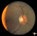
H05 Panhypoplasia | Right eye. Distinctive septo-optic dysplasia.Hypoplasia of the optic nerve. Left eye normal. Amblyopic right eye. 24 year old woman. Anatomy: Optic disc. Pathology: Hypoplasia of the optic nerve. Disease/ Diagnosis: Hypoplasia. | Image |
| 32 |
 |
H19 Panhypoplasia | Mild hypoplasia with dysplasia in right eye. Right eye. Normal left eye. Same patient as H_20. Anatomy: Optic disc. Pathology: Hypoplasia of the optic nerve. Disease/ Diagnosis: Hypoplasia. | Image |
| 33 |
 |
H20 Panhypoplasia | Mild hypoplasia with dysplasia in right eye. Left eye. Same patient as H_19. Anatomy: Optic disc. Pathology: Hypoplasia of the optic nerve. Disease/ Diagnosis: Hypoplasia. | Image |
| 34 |
 |
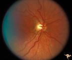
H52 Superior Segmental Optic Hypoplasia (SSOH) Topless Disc Syndrome | High exit point of central retinal vessels. Superior choroidal crescent. Complete loss of nerve fiber layer entering disc from above. Inferior altitudinal field defect. Type 1 diabetic mother. Anatomy: Optic disc. Pathology: Superior segmental optic hypoplasia (SSOH). Disease/ Diagnosis: Congenital ... | Image |
| 35 |
 |
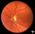
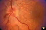
H62 Superior Segmental Optic Hypoplasia (SSOH) Topless Disc Syndrome | Right eye. Disc looks almost normal but superior nerve fiber layer is thinned and represents a mild form of SSOH. Same patient as H_61 and H_63. Anatomy: Optic disc. Pathology: Superior segmental optic hypoplasia (SSOH). Disease/ Diagnosis: Congenital anomaly. | Image |
| 36 |
 |
H80 Chiasmal Hemioptic Hypoplasia | Discs show striking nasal hypoplasia and band atrophy. DeMorsier synrome. Congenital bitemporal hemianopia with see-saw nystagmus. Note vertically oral shape of these hypoplastic nerves. The CT scan showed the median bar of the chiasm in this patient is totally hypoplastic. Anatomy: Optic disc. Path... | Image |
| 37 |
 |
H85 Chiasmal Hemioptic Hypoplasia | Congenital bitemporal hemianopia with marked bi-nasal hypoplasia. Right eye. 17 year old male. Same patient as H_84. Anatomy: Optic disc. Pathology: Chiasmal hemioptic hypoplasia. Disease/ Diagnosis: Congenital anomaly involving chiasm. | Image |
| 38 |
 |
H86 Chiasmal Hemioptic Hypoplasia | Congenital bitemporal hemianopia with nasal hypoplasia. 24 year old man. Same patient as H_87. Anatomy: Optic disc. Pathology: Chiasmal hemioptic hypoplasia. Disease/ Diagnosis: Congenital anomaly involving chiasm. | Image |
| 39 |
 |
H87 Chiasmal Hemioptic Hypoplasia | Congenital bitemporal hemianopia with nasal hypoplasia. 24 year old man. Same patient as H_86. Anatomy: Optic disc. Pathology: Chiasmal hemioptic hypoplasia. Disease/ Diagnosis: Congenital anomaly involving chiasm. | Image |
| 40 |
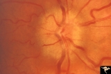 |
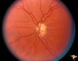
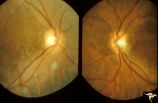
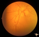
Unilateral Papilledema | Unilateral papilledema due to elevated intracranial pressure. Right eye. This eye has papilledema. Patient described transient monocular blindness with turning right eye. Asymmetric papilledema. Anatomy: Optic disc. Pathology: Unilateral papilledema. Disease/Diagnosis: Iidiopathic intracranial hyper... | Image |
| 41 |
 |
Unilateral Papilledema | Right eye. Patient had tumor on right side. Right sided large meningioma. Disc edema due to tumor. 29 year old black woman. The right disc has mild temporal pallor. Anatomy: Optic disc. Pathology: Uninaleral papilledema. Disease/Diagnosis: Meningioma of the brain. Clinical: Headache. | Image |
| 42 |
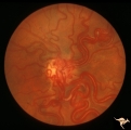 |
Vascular Disc Anomalies - Retinal Arteriovenous Malformations | Retinal arteriovenous malformations (Racemous angioma). Found in a 13 year old girl who had extension of this arteriovenous malformation up her right optic nerve into her thalamus and into her midbrain. Patient had large intra-cerebral AVM (Wyburn-Mason Syndrome). Patient died 10 years later of mass... | Image |
| 43 |
 |
Vascular Disc Anomalies - Retinal Arteriovenous Malformations | Retinal arteriovenous malformations. Two years later after interventional embolic obliteration of orbital AVM. Same patient as V_26. Ref: #73. Anatomy: Optic disc; Brain. Pathology: Arteriovenous malformation of retina and brain. Disease/Diagnosis: Wyburn-Mason syndrome. Clinical: Blindness in the i... | Image |
| 44 |
 |
Venous Anomalies - Congenital Venous Tortuosity | Congenital venous tortuosity in a young girl with a cerebral arteriovenous malformation (AVM). Same eye as V_55. This does not represent a Wyburn-Mason Syndrome. It was a congenital retinal venous anomaly, not a retinal AVM. Anatomy: Optic disc. Pathology: Congenital venous tortuosity. Disease/Diag... | Image |
| 45 |
 |
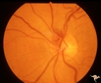
Venous Anomalies - Exit Anomalies | Disc edge veins of Kraupa in 14 year old boy. Anatomy: Optic disc. Pathology: Congenital anomaly, exit anomaly. Disease/Diagnosis: Exit anomaly, edge veins. Clinical: Asymptomatic. | Image |
| 46 |
 |
Venous Anomalies - Prepapillary Venous Convolutions (Congenital) | Sub-retinal and prepapillary venous convolutions - congenital. Edge vein. Large vein draining subretinally into the choroid. Anatomy: Optic disc. Pathology: Prepapillary venous convolutions - congenital. Disease/Diagnosis: Prepapillary venous convolutions - congenital. Clinical: Asymptomatic. | Image |
| 47 |
 |
Visible Drusen with Visual Field Loss | Left eye.16 year old girl: PP26b: buried drusen at the lower pole of the disc; PP26a: Visible drusen with visual field loss. Notice the thinning of the nerve fibers in both the superior and inferior arcuate bundles. PP26c: Goldmann visual field. Anatomy: Optic disc. Pathology: Drusen of the optic ... | Image |
| 48 |
 |
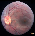
Visible Drusen with Visual Field Loss | Right eye.16 year old girl: PP26a: Visible drusen with visual field loss. Notice the thinning of the nerve fibers in both the superior and inferior arcuate bundles. PP26b: buried drusen; PP26c: Goldmann visual field. Anatomy: Optic disc. Pathology: Drusen of the optic disc. Disease/Diagnosis: Druse... | Image |
| 49 |
 |
A101 Disc Swelling due to Intraocular Hypotension | Ocular hypotension following lens replacement surgery. Retinal/macular folds. Anatomy: Optic disc. Pathology: Disc edema. Disease/ Diagnosis: Intraocular hypotension. Clinical: Low intraocular pressure or intraocular hypotension. | Image |
| 50 |
 |
H06 Panhypoplasia | Bilateral hypoplasia. Top is Right eye - moderate. Bottom is Left eye - severe. Note venous tortuosity. Good example of double ring sign. De Morsier's syndrome.Septo-optic dysplasia. Same patient as H_7. Anatomy: Optic disc. Pathology: Hypoplasia of the optic nerve. Disease/ Diagnosis: Hypoplasia. ... | Image |