Collection of materials relating to neuro-ophthalmology as part of the Neuro-Ophthalmology Virtual Education Library.
NOVEL: https://novel.utah.edu/
TO
- NOVEL720
| Title | Creator | Description | Subject | ||
|---|---|---|---|---|---|
| 451 |
 |
Papilledema 2013 | Kathleen B. Digre, MD | Objectives: What types of disc findings can be confused for papilledema List the features of true disc swelling Describe the tests you would order to evaluate and w/u papilledema List differential diagnosis of papilledema Describe possible treatments for papilledema (medical and surgical) | Papilledema |
| 452 |
 |
Optic Disc Anatomy, Variants, and Usual Discs | Kathleen B. Digre, MD | Examination of optic disc, disc anatomy, disc variation. | Optic Disc; Normal Disc Anatomy |
| 453 |
 |
Diffusion Weighted Imaging (DWI) | John Pula, MD | Diffusion weighted imaging sequences are often included as part of a routine brain MRI protocol. Imaging provides examples of DWI. | Diffusion Weighted Imaging; DWI |
| 454 |
 |
Neuromyelitis Optica (NMO) | John Pula, MD | Slideshow describing condition. | Neuromyelitis Optica; NMO |
| 455 |
 |
Vision and Alzheimer's Disease | Victoria S. Pelak, MD | Slideshow describing condition. | Alzheimer's Disease |
| 456 |
 |
Radiological Examination of the Visual System | John Pula, MD | An explanation of imaging types. | Visual System; Radiology; Imaging |
| 457 |
 |
Diffusion Tensor Imaging (DTI) | John Pula, MD | Diffusion tensor (DT) MRI applies the direction of water diffusion through tissues to map out neural pathways in the brain, such as white matter tracts. | Diffusion Tensor Imaging; DTI |
| 458 |
 |
Tonic Pupil | Adesina, Ore-Ofe, MD | PowerPoint presentation covering tonic pupil, which is damage to ciliary ganglion or short posterior ciliary nerves. It causes denervation of the ciliary body and iris sphincter muscle. | Tonic Pupil |
| 459 |
 |
Disability Evaluation Under Social Security | John Pula, MD | A. How do we evaluate visual disorders? 1. What are visual disorders? Visual disorders are abnormalities of the eye, the optic nerve, the optic tracts, or the brain that may cause a loss of visual acuity or visual fields. A loss of visual acuity limits your ability to distinguish detail, read, or do... | Visual Impairment; Visual Disorders; Legal Blindness |
| 460 |
 |
Photophobia for Patients - Large Print | Kathleen B. Digre, MD | The symptoms of light sensitivity are: an uncomfortable sense of brightness, squinting, frequent blinking, and redness of the eye (especially if the eye is dry). Involuntary eye closure and excessive blinking is seen with blepharospasm. Individuals will tend to seclude themselves in darkness. | Photophobia |
| 461 |
 |
Photophobia for Patients | Kathleen B. Digre, MD | The symptoms of light sensitivity are: an uncomfortable sense of brightness, squinting, frequent blinking, and redness of the eye (especially if the eye is dry). Involuntary eye closure and excessive blinking is seen with blepharospasm. Individuals will tend to seclude themselves in darkness. | Photophobia |
| 462 |
 |
Joint Commission International Accreditation Standards for Ambulatory Care | Joint Commission International Accreditation (JCIA) | This second edition of the Joint Commission International Accreditation Standards for Ambulatory Care contains all the standards, intent statements, and measurable elements of standards; accreditation policies and procedures; a glossary of key terms; and an index. | Standards for Ambulatory Care; JCIA |
| 463 |
 |
Facts About Ambulatory Care Accreditation | Joint Commission on Accreditation of Healthcare Organizations (JCAHO) | The Joint Commission's Ambulatory Care Accreditation Program was established in 1975, and today more than 2,000 freestanding ambulatory care organizations are Joint Commission-accredited. These organizations generally fall into the broad categories of surgical, medical/dental and diagnostic/therapeu... | Ambulatory Care Accreditation |
| 464 |
 |
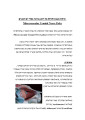
Microvascular Cranial Nerve Palsy | NANOS | Microvascular cranial nerve palsy is one of the most common causes of double vision in the older poulation. They are often referred to as "diabetic" palsies. They will resolve without leaving any double vision. | Microvascular Cranial Nerve Palsy; Patient Brochure |
| 465 |
 |
Microvascular Cranial Nerve Palsy (Spanish) | NANOS | Microvascular cranial nerve palsy is one of the most common causes of double vision in the older poulation. They are often referred to as "diabetic" palsies. They will resolve without leaving any double vision. | Microvascular Cranial Nerve Palsy; Patient Brochure |
| 466 |
 |
Microvascular Cranial Nerve Palsy (French) | NANOS | Microvascular cranial nerve palsy is one of the most common causes of double vision in the older poulation. They are often referred to as "diabetic" palsies. They will resolve without leaving any double vision. | Microvascular Cranial Nerve Palsy; Patient Brochure |
| 467 |
 |
Microvascular Cranial Nerve Palsy (German) | NANOS | Microvascular cranial nerve palsy is one of the most common causes of double vision in the older poulation. They are often referred to as "diabetic" palsies. They will resolve without leaving any double vision. | Microvascular Cranial Nerve Palsy; Patient Brochure |
| 468 |
 |
Microvascular Cranial Nerve Palsy (Hebrew) | NANOS | Microvascular cranial nerve palsy is one of the most common causes of double vision in the older poulation. They are often referred to as "diabetic" palsies. They will resolve without leaving any double vision. | Microvascular Cranial Nerve Palsy; Patient Brochure |
| 469 |
 |
Evolution of Optociliary Veins with Perioptic Nerve Sheath | William F. Hoyt, PhD | Series of images showing progression of disc swelling and macular degeneration. Pathology: Optociliary Vein. Disease/Diagnosis: Perioptic nerve sheath meningioma evolution. Clinical notes: Visual Loss. | Optic Disc Atrophy with Special Features; Optociliary Veins; Shunt Vessels (Meningioma) |
| 470 |
 |
Crowded Disc - Family | William F. Hoyt, PhD | Left eye. PP3 a & b: sister; PP4 a&b: brother; Congenital disc margin blurring with crowded discs. Excellent example of pseudo papilledema. Pathology: Normal variation of the optic disc. Disease/Diagnosis: Normal variation of the optic disc. Crowded disc. Clinical notes: Appearance due to too many f... | Pseudopapilledema; Congenital Blurred Disc |
| 471 |
 |
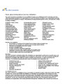
Systemic Disorders With Optic Nerve and Retinal Findings | AAO/NANOS - American Academy of Ophthalmology / North American Neuro-Ophthalmology Society | A 42-year old woman presented with a history of severe brow pain and 4 days of progressive visual loss OD. There was no increased pain on ocular rotation. Aside from heavy menses, she denied any significant past medical history. Her examination revealed acuity NLP OD, 20/25 OS; color vision 9/10 OS;... | Syphilis |
| 472 |
 |
Acute Multifocal Pigment Epithelium Epitheliopathy (AMPEE) | Gregory P. Van Stavern, MD | Images providing example of Acute Multifocal Pigment Epithelium Epitheliopathy (AMPEE) | Acute Multifocal Pigment Epithelium Epitheliopathy (AMPEE) |
| 473 |
 |
Histoplasmosis | Gregory P. Van Stavern, MD | Histoplasmosis, a fungus, can present acutely as a systemic condition. This image shows signs of Histoplasmosis. | Histoplasmosis |
| 474 |
 |
Multifocal Choroiditis | Gregory P. Van Stavern, MD | Multi-focal choroiditis is usually a bilateral choroidopathy seen more frequently in women associated with punched out appearing lesions occasionally with pigment around the edges. Image provides example. | Multi-Focal Choroiditis Panuveitis |
| 475 |
 |
Retinitis Pigmentosa | Gregory P. Van Stavern, MD | Retinitis pigmentosa is a retinal/choroidal degeneration caused by various genetic defects. The term retinitis pigmentosa is really a misnomer since it is not inflammation (retinitis) and it is not a disease of the pigmentary system (pigmentosa). | Retinitis Pigmentosa |