Best known for his world-renowned neuro-ophthalmology unit based at the University of California, San Francisco, William Hoyt, MD collected here more than 850 of his best images covering a wide range of disorders.
William F. Hoyt, MD, Professor Emeritus of Ophthalmology, Neurology and Neurosurgery, Department of Ophthalmology, University of California, San Francisco.
NOVEL: https://novel.utah.edu/
TO
| Title | Description | Type | ||
|---|---|---|---|---|
| 351 |
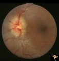 |
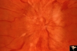
C303 Nodular Papillopathies (Sarcoid) | Lumpy infiltrative papillopathy in a patient with proven sarcoid. Anatomy: Optic disc. Pathology: Axoplasmic stasis due to sarcoid infiltration. Disease/ Diagnosis: Sarcoid papillopathy. Clinical: Unknown? | Image |
| 352 |
 |
C304 Nodular Papillopathies (Sarcoid) | Lumpy nodular disc infiltration from sarcoid. Anatomy: Optic disc. Pathology: Axoplasmic stasis due to sarcoid infiltration. Disease/ Diagnosis: Sarcoid papillopathy. Clinical: Unknown? | Image |
| 353 |
 |
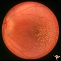
C308 Nodular Papillopathies (Sarcoid) | Nodular infiltrative papillopathy in a patient with sarcoid. Woman. Anatomy: Optic disc. Pathology: Axoplasmic stasis due to sarcoid infiltration. Disease/ Diagnosis: Sarcoid papillopahty. Clinical: Unknown? | Image |
| 354 |
 |
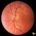
D202 Disc Edema with Systemic Hypertension | Right eye. Note generalized arteriole narrowing. Low grade disc edema and multiple splinter hemorrhages. The patient had severe hypertension from kidney failure. Additional yellow intraretinal exudate at the macula. 20 year old male patient. Pair with D2_01. Anatomy: Optic disc; Retina; Retinal art... | Image |
| 355 |
 |
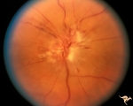
C403 Luetic Papillopathy (Gumma of the Optic Disc) | 40 year old man with AIDS and neurosyphillis with severe visual field defect. The disc is pale and swollen and its arteries are strikingly narrowed (syphillitic vasculitis). Anatomy: Optic disc. Pathology: Axoplasmic stasis due to syphillitic infection. Disease/ Diagnosis: Luetic papillopathy (Syphy... | Image |
| 356 |
 |
C206 Papillitis with Macular Star, Cat Scratch Disease | Proven Bartonella neuroretinitis. Resolved papillitis with residual retinal exudate. Man. Anatomy: Optic disc; Retina. Pathology: Axoplasmic stasis due to inflammation. Disease/ Diagnosis: Bartonella Henslae (Cat Scratch). Clinical: Visual blurring; Ocular edema with macular star (ODEMS). | Image |
| 357 |
 |
C207 Papillitis with Macular Star, Cat Scratch Disease | Proven Bartonella neuroretinitis. Anatomy: Optic disc; Retina. Pathology: Axoplasmic stasis due to inflammation. Disease/ Diagnosis: Bilateral Bartonella Henslae (Cat Scratch). Clinical: Visual blurring; Ocular disc edema with macular star (ODEMS). | Image |
| 358 |
 |
C402 Luetic Papillopathy (Gumma of the Optic Disc) | November 2001. Same eye as C4_01 after treatment with penicillin. Disc swelling went away and good visual function returned. Anatomy: Optic disc. Pathology: Axoplasmic stasis due to syphillitic infection. Disease/ Diagnosis: Luetic papillopathy (Syphillis). Clinical: Improving visual loss. | Image |
| 359 |
 |
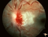
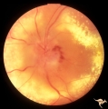
C401 Luetic Papillopathy (Gumma of the Optic Disc) | Diffuse optic disc swelling with tortuous capillary dilations indicating inflammatory cellular infiltration. October 2001. Same eye as C4_02. Anatomy: Optic disc. Pathology: Axoplasmic stasis due to syphillitic infection. Luetic papillopathy (Syphyllis). Clinical: Visual loss. | Image |
| 360 |
 |
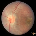
C305 Nodular Papillopathies (Sarcoid) | July 1984 shows multiple infiltrative nodules on the optic disc in addition to circumferential subretinal yellow exudates. 32 year old black woman. Same patient as C3_06 and C3_07. Anatomy: Optic disc; Retina. Pathology: Axoplasmic stasis due to sarcoid infiltration and retinal exudation. Disease/ ... | Image |
| 361 |
 |
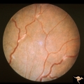
C307 Nodular Papillopathies (Sarcoid) | Fluorescein angiogram shows striking staining of the sarcoid nodules. July 1984. Same patient as C3_05 and C3_06. Corresponds with July 1984 image, C3_06. Anatomy: Optic disc. Pathology: Axoplasmic stasis due to sarcoid infiltration. Disease/ Diagnosis: Sarcoid papillopathy. Clinical: Unknown? | Image |
| 362 |
 |
C302 Nodular Papillopathies (Sarcoid) | Perivenous Inflammatory Cuffing in a Patient with Proven Sarcoid. Left eye. Pair with C3_01. Anatomy: Retina. Pathology: Axoplasmic stasis due to sarcoid infiltration with retinal venous exudation? Disease/ Diagnosis: Sarcoid papillopathy with perivenous inflammatory disease. Clinical: Unknown? | Image |
| 363 |
 |
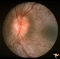
C306 Nodular Papillopathies (Sarcoid) | Lumpy disc swelling with retinal folds and a macular star in a patient with sarcoid. Presentation in October 1983. Same patient as C3_05 and C3_07. Anatomy: Optic disc; Retina. Pathology: Axoplasmic stasis due to sarcoid infiltration. Disease/ Diagnosis: Axoplasmic stasis due to sarcoid infiltration... | Image |
| 364 |
 |
C301 Nodular Papillopathies (Sarcoid) | Disc swelling. Sarcoid papillopathy. Note infiltrative nodule at 9:00 on the disc.The patient had proven sarcoid. Perivenous inflammatory cuffing visible on image C3_02. Right eye. Pair with C3_02. Anatomy: Optic disc; Retina. Pathology: Axoplasmic stasis due to sarcoid infiltration. Disease/ Diagn... | Image |
| 365 |
 |
C113 Papillitis, Retrobulbar Neuritis | Woman with herpes. Acute retinal necrosis with papillitis an arcuate neuro-retinitis. Right eye. Notice the large arcuate defect extending fromt he disc to the retina of retinal necrosis. Pair with C1_12. Anatomy: Optic disc; Retina. Pathology: Axoplasmic stasis due to inflammation; Retinal necrosis... | Image |
| 366 |
 |
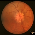
C115 Papillitis, Retrobulbar Neuritis | Demyelinative optic neuropathy with mild disc swelling. This eye had a large central scotoma. Note the bland disc margin swelling from 2:00 to 4:00. This swelling constitutes spill over edema from the main focus of the neuritis which lies behind the eyeball. Visual acuity was 2200. Anatomy: Optic di... | Image |
| 367 |
 |
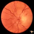
C114 Papillitis, Retrobulbar Neuritis | Epstien-Barr Virus papillitis with remarkably good recovery. Steroid responsive. Woman. Anatomy: Optic disc. Pathology: Axoplasmic stasis due to inflammation. Disease/ Diagnosis: Epstein Barr Virus with papillitis. Clinical: Visual loss that was steroid responsive. | Image |
| 368 |
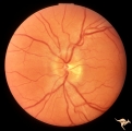 |
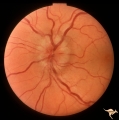
C116 Papillitis, Retrobulbar Neuritis | Demyelinative optic neuropathy with mild disc swelling. Note the mild disc margin blurring. The main focus of the neuritis lies behind the eye. 20/20 visual acuity. Anatomy: Optic disc; Optic nerve. Pathology: Axoplasmic stasis due to inflammation; Acute demyelination. Disease/ Diagnosis: Acute sub-... | Image |
| 369 |
 |
C205 Papillitis with Macular Star, Cat Scratch Disease | Proven Bartonella neuroretinitis. Macular star present, but not visible on image. 33 year old woman. Anatomy: Optic disc; Retina. Pathology: Axoplasmic stasis due to inflammation. Disease/ Diagnosis: Bartonella Henslae (Cat Scratch). Clinical: Visual blurring. | Image |
| 370 |
 |
C201 Papillitis with Macular Star, Cat Scratch Disease | Proven Bartonella neuroretinitis. Notice the deposit of exudates of Henle's layer making an almost complete macular star. Anatomy: Optic disc; Retina. Pathology: Neuroretinitis; Axoplasmic stasis due to inflammation. Disease/ Diagnosis: Neuroretinitis due to Bartonella Henslae. Clinical: Visual blur... | Image |
| 371 |
 |
B407 Disc Swelling, Radiation Papillopathy | Man with blind eye. June 1985. Same patient as B401 and B402. Note the striking peripapillary intraretinal exudatation occurring at a slight distance from the disc. Anatomy: Optic disc. Pathology: Axoplasmic stasis due to ischemia; Ischemic infarction. Disease/ Diagnosis: Radiation papillopathy; Opt... | Image |
| 372 |
 |
B402 Disc Swelling, Radiation Papillopathy | Radiation papillopathy with arterial narrowing, exudation and venous dilation in man with blind eye. May 1985. Same patient as B401, B407. Anatomy: Optic disc. Pathology: Axoplasmic stasis due to ischemia. Disease/ Diagnosis: Radiation papillopahty; optic neuropathy. Clinical: Visual loss after radi... | Image |
| 373 |
 |
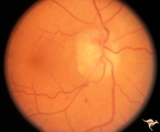
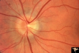
C106 Papillitis, Retrobulbar Neuritis | Papillitis with recovery of vision. Woman acupuncturist. Anatomy: Optic disc. Pathology: Axoplasmic stasis after inflammation. Disease/ Diagnosis: Optic neuritis/optic papillitis. Clinical: Visual loss with recovery. | Image |
| 374 |
 |
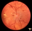
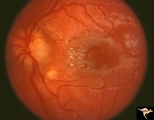
C104 Papillitis, Retrobulbar Neuritis | Post infectious papillitis with macular exudate. Anatomy: Optic disc macula. Pathology: Axoplasmic stasis due to inflammation with lipid deposit in Henle's layer. Disease/ Diagnosis: Post infectious papillitis/optic neuritis. Clinical: Visual loss after infection. | Image |
| 375 |
 |
C107 Papillitis, Retrobulbar Neuritis | Man with bilateral papillitis. Right eye. Pair with C1_08. Cause unknown. Visual field showed central scotomas. Anatomy: Optic disc. Pathology: Axoplasmic stasis due to inflammation. Disease/ Diagnosis: Neuritis of the optic nerve. Clinical: Visual loss. | Image |