Best known for his world-renowned neuro-ophthalmology unit based at the University of California, San Francisco, William Hoyt, MD collected here more than 850 of his best images covering a wide range of disorders.
William F. Hoyt, MD, Professor Emeritus of Ophthalmology, Neurology and Neurosurgery, Department of Ophthalmology, University of California, San Francisco.
NOVEL: https://novel.utah.edu/
TO
| Title | Description | Type | ||
|---|---|---|---|---|
| 326 |
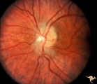 |
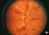
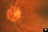
F202 Optic Nerve Sheath Meningioma | Optic nerve sheath meningioma. Note optociliary vein at 3:00. The disc is atrophic. Anatomy: Optic disc. Pathology: Chronic optic disc swelling caused by optic nerve sheath meningioma. Disease/ Diagnosis: Chronic optic disc swelling caused by optic nerve sheath meningioma. | Image |
| 327 |
 |
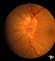
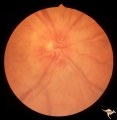
F2b11 Optic Disc Swelling from Optic Glioma | Optic disc swelling from optic glioma. Note the signs of vein occlusion and the optociliary bypass vien at 4:00. Left eye. Anatomy: Optic disc. Pathology: Optic nerve glioma. Disease/ Diagnosis: Optic nerve swelling secondary to retrobulbar optic glioma. | Image |
| 328 |
 |
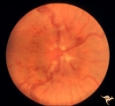
F108 Acute Disc Swelling | Chinese man with acute disc swelling. Blind in both eyes. Had large thalamic mass. (Lymphoma). Anatomy: Optic disc. Pathology: Lymphoma. Disease/ Diagnosis: Lymphoma. | Image |
| 329 |
 |
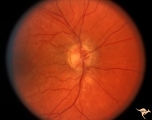
F206 Intracavernous Meningioma Extending Into the Orbit | Intracavernous meningioma extending into the orbit. Female patient. Anatomy: Optic disc. Pathology: Intracavernous meningioma. Disease/ Diagnosis: Neoplastic papillopathy. | Image |
| 330 |
 |
F204 Optic Nerve Sheath Meningioma | Optic disc swelling due to meningioma. Notice choroidal folds through the macula of left eye. Anatomy: Optic disc. Pathology: Chronic optic disc swelling caused by optic nerve sheath meningioma. Disease/ Diagnosis: Chronic optic disc swelling caused by optic nerve sheath meningioma. | Image |
| 331 |
 |
A302b Disc Swelling, Chorioretinal Disease | Bad chorioretinal scars with disc swelling. Temporal extent of chorioretinal scarring. A and B are the same eye. Anatomy: Optic disc. Pathology: Unknown. | Image |
| 332 |
 |
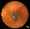
A303 Disc Swelling, Chorioretinal Disease | Neovascular net. Disc swelling with peripapillary neo-vascularization with subretinal hemorrhage. Anatomy: Optic disc; Retina. | Image |
| 333 |
 |
A301a Disc Swelling, Chorioretinal Disease | a and b same eye. Bad chorioretinal scars with disc swelling. Anatomy: Optic disc. Pathology: Unknown. | Image |
| 334 |
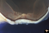 |
F2b10 Malignant Optic Nerve Glioma, Gross Pathologic Specimen | Pathologic specimen of optic nerve glioma shown in slide F2b_09. White material on top of swollen disc is myelin. Reference: Hoyt WF, Meshel LG, Lessell S, Schatz NJ, Suckling RD. Malignant optic glioma of adulthood. Brain. 1973;96(1):121-32. Anatomy: Optic disc. Pathology: Optic nerve glioma. Disea... | Image |
| 335 |
 |
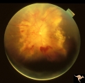
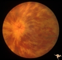
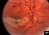
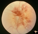
Bilateral Papilledema with Exudative Retinopathy | Bilateral Papilledema with exudative retinopathy from vitamin A toxicity. Young boy. Near blind. Anatomy: Optic disc; Retina. Pathology: Bilateral papilledema; exudative retinopathy. Disease/Diagnosis: Hypervitaminosis A causing blindness. Clinical notes: Nearly blind; Headache. | Image |
| 336 |
 |
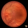
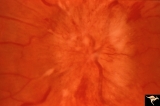
Bilateral Papilledema with Exudative Retinopathy | Left eye. Bilateral Papilledema with exudative retinopathy from vitamin A toxicity. Young boy. Near blind. Anatomy: Optic disc; Retina. Pathology: Bilateral papilledema; exudative retinopathy. Disease/Diagnosis: Hypervitaminosis A causing blindness. Clinical notes: Nearly blind; Headache. | Image |
| 337 |
 |
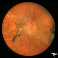
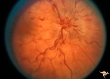
E02 Disc Swelling with Central Vein Occlusion | 37 year old black male with sickle cell C causing unilateral central retinal vien occlusion. Anatomy: Optic disc; Retina. Pathology: Occlusion of the central retinal vein. Disease/ Diagnosis: Disc swelling due to central retial vein occlusion. Clinical: Visual blurring. | Image |
| 338 |
 |
E10 Disc Swelling with Central Vein Occlusion | Cilioretinal artery infarction after a central retinal vein occlusion. Anatomy: Optic disc; Retina. Pathology: Central retinal vein occlusion. Disease/ Diagnosis: Disc swelling due to central retinal vein occlusion. | Image |
| 339 |
 |
E04 Disc Swelling with Central Retinal Vein Occlusion | Acute CRVO, right eye with disc swelling. Male patient. Same patient as E05. Anatomy: Optic disc; Retina. Pathology: Central retinal vein occlusion. Disease/ Diagnosis: Disc swelling due to central retinal vein occlusion. Clinical: Visual blurring. | Image |
| 340 |
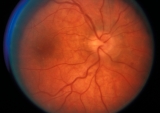 |
E05 Disc Swelling with Central Retinal Vein Occlusion | Resolving CVRO, right eye. Two months following slide E04. Male patient. Anatomy: Optic disc; Retina. Pathology: Central retinal vein occlusion. Disease/ Diagnosis: Resolved disc swelling after central retinal vein occlusion. Clinical: No symptoms. | Image |
| 341 |
 |
D201 Disc Edema with Systemic Hypertension | Left eye. Note generalized arterial narrowing. Low grade disc edema and multiple splinter hemorrhages. The patient had severe hypertension from kidney failure. Additional yellow intraretinal exudate at the macula. 20 year old male patient. Right eye. Pair with D2_02. Anatomy: Optic disc; Retina; Ret... | Image |
| 342 |
 |
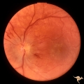
E03 Disc Swelling with Central Retinal Vein Occlusion | 36 year old woman with visual obscurations of right eye. Early CRVO, papillophlebitis. Steroid responsive. Anatomy: Optic disc; Retina. Pathology: Central retinal vein occlusion. Disease/ Diagnosis: Disc swelling due to central retinal vein occlusion. Clinical: Decreased vision in right eye. Acuity ... | Image |
| 343 |
 |
E08 Disc Swelling with Central Vein Occlusion | Pituitary adenoma with right chronic CRVO with optociliary bypass vessels. Anatomy: Optic disc; Retina. Pathology: Central retinal vein occlusion. Disease/ Diagnosis: Disc swelling due to central retinal vein occlusion. | Image |
| 344 |
 |
E09 Disc Swelling with Central Vein Occlusion | Chronic disc swelling due to CRVO. Anatomy: Optic disc; Retina. Pathology: Central retinal vein occlusion. Disease/ Diagnosis: Disc swelling due to central retinal vein occlusion. | Image |
| 345 |
 |
E11 Disc Swelling with Central Vein Occlusion | Old retinal vein occlusion with optociliary bypass vessel at 3:00. Right eye. Anatomy: Optic disc; Retina. Pathology: Central retinal vein occlusion. Disease/ Diagnosis: Disc swelling due to central retinal vein occlusion. | Image |
| 346 |
 |
E12 Disc Swelling with Central Vein Occlusion | 2nd attack of papillophlebitis. There is an optociliary bypass vessel at 4:00. Anatomy: Optic disc; Retina. Pathology: Central retinal vein occlusion. Disease/ Diagnosis: Disc swelling due to central retinal vein occlusion. | Image |
| 347 |
 |
E07 Disc Swelling with Central Vein Occlusion | 24 year old male. Papillophlebitis (CRVO) with optic disc edema. Right eye. Anatomy: Optic disc; Retina. Pathology: Central retinal vein occlusion. Disease/ Diagnosis: Disc swelling due to central retinal vein occlusion. Clinical: ??Branch retinal artery occlusion [sic]. | Image |
| 348 |
 |
E06 Disc Swelling with Central Retinal Vein Occlusion | Acute disc swelling one week after onset of symptoms. Anatomy: Optic disc; Retina. Pathology: Central retinal vein occlusion. Disease/ Diagnosis: Disc swelling due to central retinal vein occlusion. Clinical: Visual blurring. | Image |
| 349 |
 |
C402 Luetic Papillopathy (Gumma of the Optic Disc) | November 2001. Same eye as C4_01 after treatment with penicillin. Disc swelling went away and good visual function returned. Anatomy: Optic disc. Pathology: Axoplasmic stasis due to syphillitic infection. Disease/ Diagnosis: Luetic papillopathy (Syphillis). Clinical: Improving visual loss. | Image |
| 350 |
 |
C401 Luetic Papillopathy (Gumma of the Optic Disc) | Diffuse optic disc swelling with tortuous capillary dilations indicating inflammatory cellular infiltration. October 2001. Same eye as C4_02. Anatomy: Optic disc. Pathology: Axoplasmic stasis due to syphillitic infection. Luetic papillopathy (Syphyllis). Clinical: Visual loss. | Image |