AAO-NANOS Neuro-Ophthalmology Clinical Collection: Derived from the AAO-NANOS Clinical Neuro-Ophthalmology collection produced on CD. The images are of selected cases from the NANOS teaching slide exchange, and the CD was produced under the direction of Larry Frohman, MD and Andrew Lee, MD.
The American Academy of Ophthalmology (AAO); The North American Neuro-Ophthalmology Association (NANOS).
NOVEL: https://novel.utah.edu/
TO
| Title | Creator | Description | ||
|---|---|---|---|---|
| 326 |
 |
Neuro-Ophthalmic Consequences of Therapy | Mark J. Kupersmith, MD | radiation retinopathy may mimic diabetic or hypertensive optic neuropathy. A history of irradiation to the eye, orbit, or head is mandatory. Radiation retinopathy usually occurs many months after radiation therapy. |
| 327 |
 |
Neuro-Ophthalmic Consequences of Therapy | Mark J. Kupersmith, MD | radiation retinopathy may mimic diabetic or hypertensive optic neuropathy. A history of irradiation to the eye, orbit, or head is mandatory. Radiation retinopathy usually occurs many months after radiation therapy. |
| 328 |
 |
Systemic Disorders With Optic Nerve and Retinal Findings | Larry P. Frohman, MD | A 29-year-old African American woman presented with headaches, bilateral transient visual obscurations, blurred vision, numbness, and weakness of the lower extremities with myalgia and joint pains. She had an unplanned 12-pound weight loss over 2 months. A neurologist and internist diagnosed her wit... |
| 329 |
 |
Systemic Disorders With Optic Nerve and Retinal Findings | Larry P. Frohman, MD | A 29-year-old African American woman presented with headaches, bilateral transient visual obscurations, blurred vision, numbness, and weakness of the lower extremities with myalgia and joint pains. She had an unplanned 12-pound weight loss over 2 months. A neurologist and internist diagnosed her wit... |
| 330 |
 |
Systemic Disorders With Optic Nerve and Retinal Findings | Larry P. Frohman, MD | A 35-year-old African-American woman had gradual bilateral painless visual loss over 3 months. When initially seen, the visual acuities were HM OD, NLP OS. The MRI showed diffused enhancement of the optic nerves and lacrimal glands. The evaluation strongly suggested sarcoidosis, with elevated angiot... |
| 331 |
 |
Systemic Disorders With Optic Nerve and Retinal Findings | Larry P. Frohman, MD | A 35-year-old African-American woman had gradual bilateral painless visual loss over 3 months. When initially seen, the visual acuities were HM OD, NLP OS. The MRI showed diffused enhancement of the optic nerves and lacrimal glands. The evaluation strongly suggested sarcoidosis, with elevated angiot... |
| 332 |
 |
Chiasmal Syndromes | Larry P. Frohman, MD | This 36-year-old woman presented in 1988 with 3 weeks of vertical binocular diplopia. She was a known amblyope OD. Her examination was notable for a right hyperdeviation of 1 PD present in right gaze and a subtle left noncongruous homonymous field defect. She was lost to follow-up, but 5 years later... |
| 333 |
 |
Chiasmal Syndromes | Larry P. Frohman, MD | This 36-year-old woman presented in 1988 with 3 weeks of vertical binocular diplopia. She was a known amblyope OD. Her examination was notable for a right hyperdeviation of 1 PD present in right gaze and a subtle left noncongruous homonymous field defect. She was lost to follow-up, but 5 years later... |
| 334 |
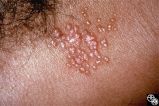 |
Systemic Disorders With Optic Nerve and Retinal Findings | Larry P. Frohman, MD | Skin rashes occur in about 30 percent of patients with sarcoid. When seen, the rashes offer an accessible site for obtaining histologic material for confirmation of the clinical diagnosis. Pair with 91_68. |
| 335 |
 |
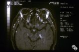
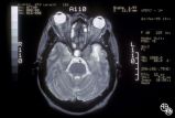
Neuro-Ophthalmic Imaging-MRI | Mitchell J. Wolin, MD | Axial view of Arnold-Chiari malformation on a patient with downbeat nystagmus. Note the presence of the cerebellar tonsils posterior to the caudal medulla. In addition to downbeat nystagmus, Arnold-Chiari malformations can sometimes lead to increased intracranial pressure and papilledema. |
| 336 |
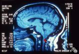 |
Neuro-Ophthalmic Imaging-MRI | Mitchell J. Wolin, MD | Sagittal view of Arnold-Chiari malformation on a patient with downbeat nystagmus. The compression of the cervicomedullary junction is clearly depicted in the sagittal view. |
| 337 |
 |
Ocular Manifestations of Congenital/Inherited Diseases | William Fletcher Hoyt, MD | Neurofibromatosis, type 1, is an autosomal dominant phakomatosis characterized by Lisch nodules of the iris (hamartomas) plexiform neurofibromas, café-au-lait spots on the skin, and axillary freckling. Intracranial tumors such as optic pathway gliomas may occur. Disease/Diagnosis: Neurofibromatosis... |
| 338 |
 |
Ocular Manifestations of Congenital/Inherited Diseases | Larry P. Frohman, MD | On optic nerve CT scan, this patient with neurofibromatosis, type 1, shows the classic railroad-track sign of optic nerve meningioma and the kink sign of optic nerve glioma. Disease/Diagnosis: Neurofibromatosis, Type 1. |
| 339 |
 |
Isolated Optic Neuritis/Neuropathy | John A. Charley, MD | Several Primary Mutations result in Leber's hereditary optic neuropathy, including mitochondrial deletions at positions 11778, 3460, and 14484. Although the 11778 is the most common mutation, the 11484 has the best prognosis for spontaneous recovery. This case exhibits the 3460 mutation. |
| 340 |
 |
Isolated Optic Neuritis/Neuropathy | John A. Charley, MD | Several Primary Mutations result in Leber's hereditary optic neuropathy, including mitochondrial deletions at positions 11778, 3460, and 14484. Although the 11778 is the most common mutation, the 11484 has the best prognosis for spontaneous recovery. This case exhibits the 3460 mutation. |
| 341 |
 |
Isolated Optic Neuritis/Neuropathy | John A. Charley, MD | Several Primary Mutations result in Leber's hereditary optic neuropathy, including mitochondrial deletions at positions 11778, 3460, and 14484. Although the 11778 is the most common mutation, the 11484 has the best prognosis for spontaneous recovery. This case exhibits the 3460 mutation. |
| 342 |
 |
Isolated Optic Neuritis/Neuropathy | John A. Charley, MD | Several Primary Mutations result in Leber's hereditary optic neuropathy, including mitochondrial deletions at positions 11778, 3460, and 14484. Although the 11778 is the most common mutation, the 11484 has the best prognosis for spontaneous recovery. This case exhibits the 3460 mutation. |
| 343 |
 |
Isolated Optic Neuritis/Neuropathy | John A. Charley, MD | Several Primary Mutations result in Leber's hereditary optic neuropathy, including mitochondrial deletions at positions 11778, 3460, and 14484. Although the 11778 is the most common mutation, the 11484 has the best prognosis for spontaneous recovery. This case exhibits the 3460 mutation. |
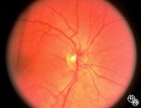
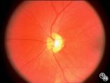
| 344 |
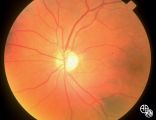 |
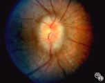
Isolated Congenital Optic Disc Anomalies | Roger Turbin, MD | Shown are the fundi of a - year old child with dominant optic atrophy, 20/200 OU. Pair with 1996_59. Disease/Diagnosis: Congenital optic atrophy. |
| 345 |
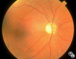 |
Isolated Congenital Optic Disc Anomalies | Roger Turbin, MD | Shown are the fundi of a - year old child with dominant optic atrophy, 20/200 OU. Pair with 1996_60. Disease/Diagnosis: Congenital optic atrophy. |
| 346 |
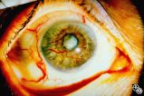 |
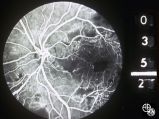
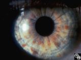
Neuro-Ophthalmic Vascular Disease | Mark L. Moster, MD | Neovascularization of the iris may form in response to an ischemic disease of the retina, such as diabetic retinopathy. Carotid artery occlusion may result in ocular ischemia that may induce neovascularization.This is a dramatic image of iris neovascularization. |
| 347 |
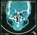 |
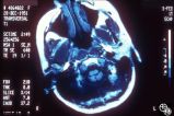
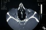
Neuro-Ophthalmic Imaging-CT Scan | Mitchell J. Wolin, MD | This is a patient with trauma leading to enucleation, with swelling years later over the implant. This is a presumed chronic abscess between orbit and dura. |
| 348 |
 |
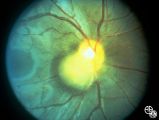
Ocular Manifestations of Congenital/Inherited Diseases | William Fletcher Hoyt, MD | Tuberous sclerosis is an autosomal dominant phakomatosis characterized by astrocytic hamartomas of the retina or optic nerve, adenoma sebaceum of the face, periungual fibromas, and astrocytic hamartomas of the brain, with secondary seizures and mental retardation. Disease/Diagnosis: Tuberous Scleros... |
| 349 |
 |
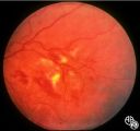
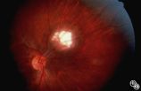
Ocular Manifestations of Congenital/Inherited Diseases | Anthony C. Arnold, MD | Note the retinal/optic nerve head astrocytoma in a 12-year-old boy with tuberous sclerosis. Disease/Diagnosis: Tuberous Sclerosis. |
| 350 |
 |
Neuro-Ophthalmic Vascular Disease | Mark J. Kupersmith, MD | Aneurysms of the intracranial circulation may act as mass lesions and compress the afferent of efferent visual pathway. Ophthalmic artery aneurysms may compress the optic nerve and result in an optic neuropathy (ie, visual loss, afferent pupillary defect, optic atrophy). Treatment includes endovascu... |