Collection of materials relating to neuro-ophthalmology as part of the Neuro-Ophthalmology Virtual Education Library.
NOVEL: https://novel.utah.edu/
TO
- NOVEL730
| Title | Creator | Description | Subject | ||
|---|---|---|---|---|---|
| 301 |
 |
Pituitary Tumor (Tagalog) | NANOS | Pituitary tumors are benign (non-cancerous) overgrowth of cells that make up the pituitary gland (the master gland that regulates other glands in the body). | Pituitary Tumor; Patient Brochure |
| 302 |
 |
TAO (Danish) | NANOS | This is an autoimmune condition where your body's immune system is producing factors that stimulate enlargement of the muscles that move the eye. | Thyroid Eye Disease; Patient Brochure |
| 303 |
 |
Hemifacial Spasm (Korean) | NANOS | Involuntary contractions, called "spasms," of the muscles on one side of the face. The affected side of the face seems to "scrunch up" while the other side of the face remains normal. | Hemifacial Spasm; Patient Brochure |
| 304 |
 |
Mikrovaskulr Parese - Microvascular Cranial Nerve Palsy (Danish) | NANOS | Microvascular cranial nerve palsy is one of the most common causes of double vision in the older poulation. They are often referred to as "diabetic" palsies. They will resolve without leaving any double vision. | Microvascular CNP; Patient Brochure |
| 305 |
 |
Optic Neuritis (Tagalog) | NANOS | In the most common form of optic neuritis, the optic nerve has been attacked by the body's overactive immune system and results in decreased vision. | Optic Neuritis; Patient Brochure |
| 306 |
 |
Neuromyelitis Optica | Omar Ozgur, MD; Rudrani Banik, MD | Power point of case presentation of female with bilateral, sequential atypical optic neuritis. MRI Brain normal with no demyelination; MRI Spine shows enhancement at multiple levels and NMO antibody positive, confirming diagnosis of neuromyelitis optica (NMO). History of NMO discussed, diagnostic c... | Neuromyelitis Optica; Atypical Optic Neuritis; MRI; Plasmapheresis |
| 307 |
 |
Oculopharyngeal Muscular Dystrophy (OPMD) | Natasha Nayak, MD; Rudrani Banik, MD | Power point of case presentation of chronic, progressive ophthalmoplegia and bilateral ptosis in adult male with positive family history of similar ocular findings. Differential diagnosis with associated findings reviewed. Work up done: EMG testing consistent with myopathy. Genetic testing positiv... | Ophthalmoplegia;, Ptosis; Oculopharygneal Muscular Dystrophy; Genetic Disorder |
| 308 |
 |
Retinal Causes of a Neurologic-Type Visual Field Defect | Omar Ozgur, MD; Rudrani Banik, MD | Power point of case presentation of 47 year old female with history of breast cancer with new onset temporal visual field defect and photopsias. Differential diagnosis of homonymous hemianopia discussed; retinal causes of neurologic-type visual field defects reviewed including: white dot syndrome (m... | Homonymous Hemianopia; Neurologic Visual Field Defect; Temporal Visual Field Defect; White Dot Syndrome; Multiple Evanescent White Dot Syndrome (MEWDS); Cancer-Associated Retinopathy; Tamoxifen Retinopathy; Autoimmune Retinopathy |
| 309 |
 |
Palinopsia: Some Visions Never Fade | Amrita-Amanda D. Lakraj, MD; Ryan D. Walsh, MD | This is a PowerPoint presentation, which teaches the symptom of palinopsia through a video of a patient's chief complaint in which he describes the symptom almost according to a textbook. This video is followed by a brief explanation of the etiology, management, and importance of diagnosing this sym... | Palinopsia; Visual Disturbance; Ghosting |
| 310 |
 |
Direct Carotid Cavernous Fistula | Emory Eye Center | Slideshow describing condition. | Fistula |
| 311 |
 |
Prolactinoma in Pregnancy | Timothy Sullivan, MD; Rudrani Banik, MD | Power point of case of prolactinoma which became symptomatic during pregnancy with visual field loss. Discussion of prolactinomas and their management. Patient underwent observation only. Post-partum examination revealed resolution of bitemporal field defect with reduction in size of prolactinoma ... | Prolactinoma; Pregnancy; Bitemporal Defect |
| 312 |
 |
Pituitary Apoplexy and Hemifield Slide Phenomenon | Helen H. Yeung, MD; Rudrani Banik, MD | PowerPoint of case presentation of pituitary apoplexy. Patient presented with bilateral severe visual loss and bilateral ophthalmoplegia from partial third nerve palsies (pupil-sparing with no ptosis) from midbrain compression. After transsphenoidal surgery with decompression of mass and steroids, ... | Pituitary Apoplexy; Hemifield Slide; Bitemporal Defect; Partial Third Nerve Palsy |
| 313 |
 |
Wallenberg Syndrome and Skew Deviation | Lauren Schneider, MD; Rudrani Banik, MD | Power point of case presentation of acute Wallenberg Syndrome associated with vertical diplopia, found by 3 step and supine testing to be consistent with skew deviation. | Wallenberg Syndrome; Skew Deviation; Vertical Diplopia |
| 314 |
 |
Pseudotumor cerebri and Chiari Malformation | Nicole Scripsema, MD; Rudrani Banik, MD | Power point of case presentation of pseudotumor cerebri with co-existing Chiari malformation. Management of severe visual loss associated with chronic papilledema discussed, as well as possible relationship between raised intracranial pressure from pseudotumor cerebri and Chiari malformation. | Pseudotumor Cerebri; Papilledema; Chiari Malformation |
| 315 |
 |
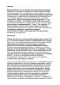
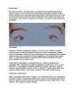
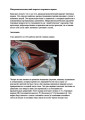
Lemierre Syndrome - A Neuroophthalmological Approach | Vinzenz A. C. Vadasz, MD; Christina Gerth-Kahlert, MD | Case report of a twenty-two year old woman with double vision after tonsillitis, caused through multiples thrombosis by an infection with fusobacterium necrophorum known as the Lemierre-Syndrome. Fig. 1: Ocular motility at ICU (lying position) Fig. 2: white arrows show thrombosis of the right opht... | Lemierre-Syndrome; Fusobacterium Necrophorum; Septic Thrombosis |
| 316 |
 |
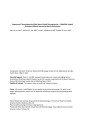
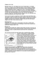
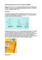
Acute Zonal Occult Outer Retinopathy (AZOOR) versus Multiple Evanescent White Dot Syndrome (MEWDS) | Asim V. Farooq, MD; Michael T. Andreoli, MD; Heather E. Moss, MD | PPT case report on acute zonal occult outer retinopathy (AZOOR) versus multiple evanescent white dot syndrome (MEWDS). | AZOOR; MEWDS; Paracentral Scotoma; Goldmann Visual Field; Photoreceptor Loss |
| 317 |
 |
Idiopathic Bilateral Neuroretinitis in a Child | Asim V. Farooq, MD; Michael T. Andreoli, MD; Molly Gilbert, MD; Heather E. Moss, MD | PPT case describing idiopathic bilateral neuroretinitis in a child. | Neuroretinitis; Pediatric; Idiopathic; Optic Atrophy |
| 318 |
 |
Superonasal Transconjunctival Optic Nerve Sheath Decompression: A Modified Surgical Technique Without Extraocular Muscle Disinsertion | Kevin E. Lai, MD; Kenneth C. Lao, MD; Peter L. Hildebrand, MD; Bradley K. Farris, MD | Report on the surgical technique and outcomes of a modified medial transconjunctival approach to optic nerve sheath decompression (ONSD) in 15 patients. Supplemental Digital Content : Video that demonstrates the stONSD procedure. m4v: http://content.lib.utah.edu/cdm/ref/collection/EHSL-NOVEL/id/22... | Superonasal Transconjunctival Optic Nerve Sheath Decompression (ONSD); Surgical Technique |
| 319 |
 |
Dry Eye Syndrome (Russian) | NANOS | People with abnormalities of the tear film are diagnosed with "dry eyes", but some patients with "dry eyes" may not feel that their eyes are "dry". Itching, burning, a scratchy sensation, a sensation that there is sand or grit in the eye, or intermittent blurring of the vision can all be symptoms of... | Dry Eye Syndrome; Patient Brochure |
| 320 |
 |
Anterior Ischemic Optic Neuropathy AION (Russian) | NANOS | Loss of blood supply to the optic nerve results in diminished visual acuity. | Anterior Ischemic Optic Neuropathy; Patient Brochure |
| 321 |
 |
Pseudotumor Cerebri (Russian) | NANOS | This is a condition in which high pressure inside your head can cause problems with vision and headache. | Pseudotumor Cerebri; Patient Brochure |
| 322 |
 |
Migraine (Russian) | NANOS | Headache on one or both sides of the brain, and may include symptoms of nausea, vomiting, and sensitivity to light. | Migraine; Patient Brochure |
| 323 |
 |
Anisocoria (Russian) | NANOS | The pupil in the right eye and left eye are not the same size. | Anisocoria; Patient Brochure |
| 324 |
 |
Microvascular Cranial Nerve Palsy_Russian | NANOS | Microvascular cranial nerve palsy is one of the most common causes of double vision in the older poulation. They are often referred to as "diabetic" palsies. They will resolve without leaving any double vision. | Microvascular Cranial Nerve Palsy; Patient Brochure |
| 325 |
 |
Homonymous Hemianopia (Russian) | NANOS | This refers to an absence of vision towards one side of the visual world in each eye. The damage that caused this problem is in the brain and not in the eyes. | Homonymous Hemianopia; Patient Brochure |