AAO-NANOS Neuro-Ophthalmology Clinical Collection: Derived from the AAO-NANOS Clinical Neuro-Ophthalmology collection produced on CD. The images are of selected cases from the NANOS teaching slide exchange, and the CD was produced under the direction of Larry Frohman, MD and Andrew Lee, MD.
The American Academy of Ophthalmology (AAO); The North American Neuro-Ophthalmology Association (NANOS).
NOVEL: https://novel.utah.edu/
TO
Filters: Collection: "ehsl_novel_aao_nanos"
| Title | Creator | Description | ||
|---|---|---|---|---|
| 301 |
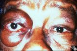 |
Ocular Manifestations of Systemic Disorders | Mitchell J. Wolin, MD | Thyroid eye disease is the most common cause of unilateral or bilateral proptosis in the adult patient. Other signs of thyroid eye disease should be sought, including lid retraction, inferior scleral show, and lid lag. Patients with markedly asymmetric or strictly unilateral proptosis should probabl... |
| 302 |
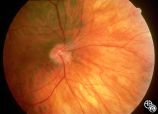 |
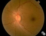
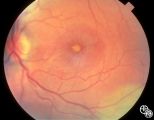
Isolated Congenital Optic Disc Anomalies | William Fletcher Hoyt, MD | This patient demonstrates bilateral tilted optic discs. Patients with this congenital optic disc anomaly may be asymptomatic or have bitemporal visual field defects that do not respect the vertical midline. Disease/Diagnosis: Tilted Discs. |
| 303 |
 |
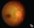
Systemic Disorders With Optic Nerve and Retinal Findings | Larry P. Frohman, MD | A 42-year old woman presented with a history of severe brow pain and 4 days of progressive visual loss OD. There was no increased pain on ocular rotation. Aside from heavy menses, she denied any significant past medical history. Her examination revealed acuity NLP OD, 20/25 OS; color vision 9/10 OS;... |
| 304 |
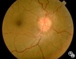 |
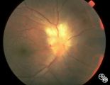
Systemic Disorders With Optic Nerve and Retinal Findings | Larry P. Frohman, MD | A 42-year old woman presented with a history of severe brow pain and 4 days of progressive visual loss OD. There was no increased pain on ocular rotation. Aside from heavy menses, she denied any significant past medical history. Her examination revealed acuity NLP OD, 20/25 OS; color vision 9/10 OS;... |
| 305 |
 |
Systemic Disorders With Optic Nerve and Retinal Findings | Larry P. Frohman, MD | This is a 32-year-old HIV-positive man with anterior uveitis, vitritis, and bilateral papillitis from syphilis. With intravenous penicillin treatment, the optic discs and vision returned to normal. |
| 306 |
 |
Chiasmal Syndromes | Larry P. Frohman, MD | This 39-year-old HIV-positive man presented in 1982 with 1 month of bilateral vision loss. His prior evaluation had included 2 CT scans, which suggested a fullness to the chiasm, and a spinal tap that showed 6 monocytes, 14 red cells, a protein of 81 mg/dl, and a glucose of 56 mg/dl, with a negative... |
| 307 |
 |
Systemic Disorders With Optic Nerve and Retinal Findings | Larry P. Frohman, MD | This 25-year-old man presented to the eye service with a history of 3 days of decreased vision OD. His past medical history was unremarkable. His examination showed acuities of 20/25 OU, with intact color plates, a 0.3 log unit of RAPD OD, and an inferior arcuate scotoma. The photos (Images 95_42, 9... |
| 308 |
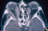 |
Ocular Manifestations of Systemic Disorders | Rosa A. Tang, MD | Thyroid eye disease may result in proptosis and restrictive external ophthalmoplegia. The extracoular muscles are often diffusely enlarged with sparing of the tendons. |
| 309 |
 |
Ocular Manifestations of Systemic Disorders | Rosa A. Tang, MD | Thyroid eye disease may cause proptosis and extraocular muscle enlargement that may be seen on orbital imaging studies. In general, coronal images allow the best visualization of the extraocular muscle enlargement. Pair with 94_45 and 94_46. |
| 310 |
 |
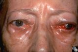
Ocular Manifestations of Systemic Disorders | Rosa A. Tang, MD | Thyroid eye disease can result in significant upper eyelid retraction and axial proptosis resulting in exposure keratopathy. |
| 311 |
 |
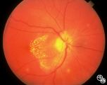
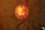
Optic Neuropathies | Michael Wall, MD | Optic disc edema with a macular star figure may occur in infectious diseases (eg, cat-scratch disease, syphilis, tuberculosis, Lyme disease), inflammatory diseases (eg, sarcoid), ischemic diseases (anterior ischemic optic neuropathy), and in papilledema. Infectious causes should be sought in patient... |
| 312 |
 |
Systemic Disorders With Optic Nerve and Retinal Findings | Joel M. Weinstein, MD | This 46-year-old woman noted progressive bilateral visual loss over a 10-month period. She had a malignant melanoma removed from her right thigh 2 years ago and excisional biopsy of an inguinal node metastasis 8 months ago. She also complained of poor night vision and rare intermittent ""sparkles"" ... |
| 313 |
 |
Systemic Disorders With Optic Nerve and Retinal Findings | Joel M. Weinstein, MD | This 46-year-old woman noted progressive bilateral visual loss over a 10-month period. She had a malignant melanoma removed from her right thigh 2 years ago and excisional biopsy of an inguinal node metastasis 8 months ago. She also complained of poor night vision and rare intermittent ""sparkles"" ... |
| 314 |
 |
Systemic Disorders With Optic Nerve and Retinal Findings | Joel M. Weinstein, MD | This 46-year-old woman noted progressive bilateral visual loss over a 10-month period. She had a malignant melanoma removed from her right thigh 2 years ago and excisional biopsy of an inguinal node metastasis 8 months ago. She also complained of poor night vision and rare intermittent ""sparkles"" ... |
| 315 |
 |
Systemic Disorders With Optic Nerve and Retinal Findings | Joel M. Weinstein, MD | This 46-year-old woman noted progressive bilateral visual loss over a 10-month period. She had a malignant melanoma removed from her right thigh 2 years ago and excisional biopsy of an inguinal node metastasis 8 months ago. She also complained of poor night vision and rare intermittent ""sparkles"" ... |
| 316 |
 |
Optic Neuropathies | Ralph A. Sawyer, MD | Optic disc edema with a macular star figure has been referred to as neuroretinitis. |
| 317 |
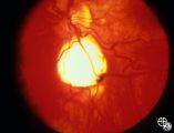 |
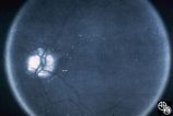
Retinal Coloboma Underneath a Relatively Normal Optic Nerve | Thomas R. Wolf, MD | Optic nerve colobomas appear as enlarged, white optic discs that are deeply excavated, often with some sapring of the superior rim. They result from an abnormal fusion of the proximal embryonic fissure. Optic nerve colobomas occur unilaterally or bilaterally with a similar frequency and can result i... |
| 318 |
 |
Isolated Congenital Optic Disc Anomalies | Thomas R. Wolf, MD | Patients with midline closure defects may exhibit abnormalities in the optic nerve, choroid, retinal pigment epithelium or retina. Anterior closure defects may result in colobomas of the structures of the anterior segment, such as the iris. Disease/Diagnosis: Coloboma. |
| 319 |
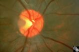 |
Isolated Congenital Optic Disc Anomalies | Anthony C. Arnold, MD | A 10-year-old girl had central visual loss due to this optic pit. Disease/Diagnosis: Optic Pit. |
| 320 |
 |
Isolated Congenital Optic Disc Anomalies | Anthony C. Arnold, MD | This is a photograph of peripheral drusen. The paired image 92_69 demonstrates the typical autofluorescence. |
| 321 |
 |
Optic Disc Drusen Autofluorescence | Anthony C. Arnold, MD | This case of optic disc drusen demonstrates the typical autofluorescence. Pair with 92_68. Imaging: Autoflourescence? |
| 322 |
 |
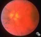
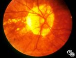
Isolated Congenital Optic Disc Anomalies | Rosa A. Tang, MD | Normally, there is no visible myelination of the nerve fiber layer in the retina. Occasionally, visible myelination occurs that takes on a characteristic white, arcuate, feathery appearance that follows the contour of the nerve fiber layer. Disease/Diagnosis: Myelinated Nerve Fiber. |
| 323 |
 |
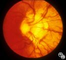
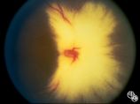
Peripapillary Staphyloma | Thomas R. Wolf, MD | Patients with ectasia of the outer layers of the eye may exhibit a posterior protrusion that appears on funduscopy as an area of deep excavation of the retina (posterior staphyloma). When it occurs around the optic disc, as in this case, it is termed a peripapillary staphyloma. This may occur in ass... |
| 324 |
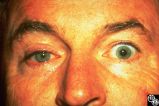 |
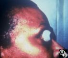
Migraine Syndrome | Mitchell J. Wolin, MD | The image shows a patient with cluster headache and eye displaying Horner's syndrome. |
| 325 |
 |
Acquired Disc Changes | Rosa A. Tang, MD | Although optociliary shunt vessels are venous collaterals that form in response to chronic venous obstruction, they may occur in patients with chronic open-angle glaucoma. |