Best known for his world-renowned neuro-ophthalmology unit based at the University of California, San Francisco, William Hoyt, MD collected here more than 850 of his best images covering a wide range of disorders.
William F. Hoyt, MD, Professor Emeritus of Ophthalmology, Neurology and Neurosurgery, Department of Ophthalmology, University of California, San Francisco.
NOVEL: https://novel.utah.edu/
TO
| Title | Description | Type | ||
|---|---|---|---|---|
| 301 |
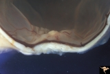 |
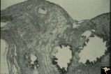
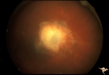
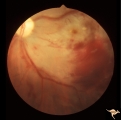
F2b10 Malignant Optic Nerve Glioma, Gross Pathologic Specimen | Pathologic specimen of optic nerve glioma shown in slide F2b_09. White material on top of swollen disc is myelin. Reference: Hoyt WF, Meshel LG, Lessell S, Schatz NJ, Suckling RD. Malignant optic glioma of adulthood. Brain. 1973;96(1):121-32. Anatomy: Optic disc. Pathology: Optic nerve glioma. Disea... | Image |
| 302 |
 |
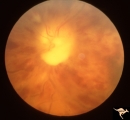
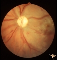
F2b11 Optic Disc Swelling from Optic Glioma | Optic disc swelling from optic glioma. Note the signs of vein occlusion and the optociliary bypass vien at 4:00. Left eye. Anatomy: Optic disc. Pathology: Optic nerve glioma. Disease/ Diagnosis: Optic nerve swelling secondary to retrobulbar optic glioma. | Image |
| 303 |
 |
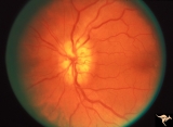
F2b13 Progression of Optic Disc Changes Caused by Malignant Optic Nerve Glioma of Adulthood | Progression. Group with F2b_12_1 and F2b_14_3. 69 year old male. April 22, 1992. There are signs of CVRO. Reference: Hoyt WF, Meshel LG, Lessell S, Schatz NJ, Suckling RD. Malignant optic glioma of adulthood. Brain. 1973;96(1):121-32. Anatomy: Optic disc. Pathology: Optic nerve glioma. Disease/ Diag... | Image |
| 304 |
 |
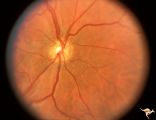
F2b14 Progression of Optic Disc Changes Caused by Malignant Optic Nerve Glioma of Adulthood | Progression. Group with F2b_12_1 and F2b_13_2. 69 year old male. Shows signs of myelin being squeezed through the optic disc into the eye. June 6, 1992. Reference: Hoyt WF, Meshel LG, Lessell S, Schatz NJ, Suckling RD. Malignant optic glioma of adulthood. Brain. 1973;96(1):121-32. Anatomy: Optic di... | Image |
| 305 |
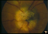 |
F401 Pigment Epithelial Hamartoma of Optic Disc | Optic disc tumor discovered incidentally in a 32 year old Asian woman who had no complaints about visual function in her involved left eye. Fundus slide shows granular elevation of left disc obscurring major disc vessels. Some of the granules has a shiny crystalline appearance. Near the vessel entra... | Image |
| 306 |
 |
F403 Pigment Epithelial Hamartoma of Optic Disc | Optic disc tumor discovered incidentally in a 32 year old Asian woman who had no complaints about visual function in her involved left eye. Fundus slide shows granular elevation of left disc obscuring major disc vessels. Some of the granules has a shiny crystalline appearance. Near the vessel entran... | Image |
| 307 |
 |
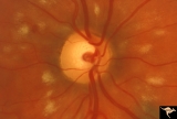
Familial Drusen | Right eye: Mother with obvious optic nerve drusen. Note the blurred temporal margin where buried drusen can not be seen.; PP_11b: mother visible drusen; Buried drusen; lumpy disc. Combine with PP_1a & b and PP_2 (sons) and PP_11c (daughter). Anatomy: Optic disc. Pathology: Drusen of the optic dis... | Image |
| 308 |
 |
Familial Drusen | Left eye. PP_11b: Mother visible drusen; buried drusen; lumpy disc. PP_11a: Mother with obvious optic nerve drusen; Combine with PP_1a & b and PP_2 (sons) and PP_11c (daughter). Anatomy: Optic disc. Pathology: Drusen of the optic disc. Disease/Diagnosis: Drusen of the optic disc. Clinical: Congen... | Image |
| 309 |
 |
Familial Drusen | PP11c: daughter: buried drusen; lumpy disc. Combine with PP1a & b and PP2 (brothers) and PP11a & b (mother). Anatomy: Optic disc. Pathology: Drusen of the optic disc. Disease/Diagnosis: Drusen of the optic disc. Clinical: Congenital dominant hereditary drusen. | Image |
| 310 |
 |
G101 Evulsion | BB injury of the optic nerve with traumatic evulsion. The missile went through the eyeball and hit the optic disc. Anatomy: Optic disc. Pathology: Optic nerve has been evulsed. Disease/ Diagnosis: Evulsion of the optic disc. | Image |
| 311 |
 |
G102 Evulsion | Partial evulsion of the left optic nerve. Anatomy: Optic disc. Pathology: Optic nerve has been evulsed. Disease/ Diagnosis: Evulsion of the optic disc. | Image |
| 312 |
 |
G103 Evulsion | Partial evulsion of the right optic nerve. Notice what is left of superior optic nerve. Anatomy: Optic disc. Pathology: Optic disc has been evulsed. Disease/ Diagnosis: Evulsion of the optic disc. | Image |
| 313 |
 |
G204 Purtchers Traumatic Retinopathy | Right eye. Blind due to chest crush with broken ribs. 18 year old male. Anatomy: Optic disc. Pathology: Varied peripapillary ischemic retinopathy. Disease/ Diagnosis: Purtchers traumatic retinopathy. | Image |
| 314 |
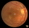 |
G205 Purtchers Traumatic Retinopathy | Right eye. Purtcher's retinopathy caused by chest crush from seat belt. Anatomy: Optic disc. Pathology: Varied peripapillary ischemic retinopathy. Disease/ Diagnosis: Purtchers traumatic retinopathy. | Image |
| 315 |
 |
G206 Purtchers Traumatic Retinopathy | Left eye. After auto accident in which the patient's chest was squeezed. Same eye as G2_07. Anatomy: Optic disc. Pathology: Varied peripapillary ischemic retinopathy. Disease/ Diagnosis: Purtchers traumatic retinopathy. | Image |
| 316 |
 |
G207 Purtchers Traumatic Retinopathy | Left eye. Large pre-retinal hemorrhage. Same eye as G2_06. Anatomy: Optic disc. Pathology: Varied peripapillary ischemic retinopathy. Disease/ Diagnosis: Purtchers traumatic retinopathy. | Image |
| 317 |
 |
G208 Traumatic AION | Traumatic vitreopapillary evulsion (traumatic AION). Traumatic AION from evulsion of the vitreopapillary adhesion. Leakage on disc surface where vitreous was adherent. Pair with G2_9b. Anatomy: Optic disc. Pathology: AION. Disease/ Diagnosis: Traumatic AION. | Image |
| 318 |
 |
G209 Traumatic AION | Traumatic vitreopapillary evulsion (traumatic AION). Fluorescein angiogram shows petal shaped avascular zones on the surface of the disc. Pair with G2_8a. Anatomy: Optic disc. Pathology: AION. Disease/ Diagnosis: Traumatic AION. Imaging: Flourescein angiogram. | Image |
| 319 |
 |
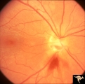
H01 Panhypoplasia | Extreme hypoplasia. Very small disc. Peri-papillary halo (choroidal). Right eye. Note: normal vessels. Same patient as H_2. Anatomy: Optic disc. Pathology: Hypoplasia of the optic nerve. Disease/ Diagnosis: Hypoplasia. Clinical: Blind child. | Image |
| 320 |
 |
H02 Panhypoplasia | Extreme hypoplasia. Very small disc. Peri-papillary halo (choroidal). Left eye. Note: normal vessels. Same patient as H_1. Anatomy: Optic disc. Pathology: Hypoplasia of the optic nerve. Disease/ Diagnosis: Hypoplasia. Clinical: Blind child. | Image |
| 321 |
 |
H03 Panhypoplasia | Extreme hypoplasia. Note absence of retinal nerve fiber layer. Left eye. Girl. Same patient as H_4. Anatomy: Optic disc. Pathology: Hypoplasia of the optic nerve. Disease/ Diagnosis: Hypoplasia. Clinical: Left eye. Girl. | Image |
| 322 |
 |
H04 Panhypoplasia | Right eye. Normal eye. Girl. Same patient as H_3. Anatomy: Optic disc. Pathology: Hypoplasia of the optic nerve. Disease/ Diagnosis: Hypoplasia. | Image |
| 323 |
 |
H05 Panhypoplasia | Right eye. Distinctive septo-optic dysplasia.Hypoplasia of the optic nerve. Left eye normal. Amblyopic right eye. 24 year old woman. Anatomy: Optic disc. Pathology: Hypoplasia of the optic nerve. Disease/ Diagnosis: Hypoplasia. | Image |
| 324 |
 |
H06 Panhypoplasia | Bilateral hypoplasia. Top is Right eye - moderate. Bottom is Left eye - severe. Note venous tortuosity. Good example of double ring sign. De Morsier's syndrome.Septo-optic dysplasia. Same patient as H_7. Anatomy: Optic disc. Pathology: Hypoplasia of the optic nerve. Disease/ Diagnosis: Hypoplasia. ... | Image |
| 325 |
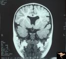 |
H07 Panhypoplasia | MRI Scan, coronal view showing absence of septum pellucidum. Hypoplastic chiasm. De Morsier's syndrome. Same patient as H_6. Anatomy: Optic disc. Pathology: Hypoplasia of the optic nerve. Disease/ Diagnosis: Hypoplasia. Imaging: MRI scan. | Image |