AAO-NANOS Neuro-Ophthalmology Clinical Collection: Derived from the AAO-NANOS Clinical Neuro-Ophthalmology collection produced on CD. The images are of selected cases from the NANOS teaching slide exchange, and the CD was produced under the direction of Larry Frohman, MD and Andrew Lee, MD.
The American Academy of Ophthalmology (AAO); The North American Neuro-Ophthalmology Association (NANOS).
NOVEL: https://novel.utah.edu/
TO
| Title | Creator | Description | ||
|---|---|---|---|---|
| 301 |
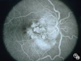 |
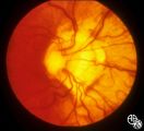
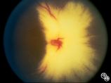
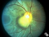
Pseudopapilledema With Subretinal Neovascularization on Fluorescein Angiogram | Joel M. Weinstein, MD | This 8-year-old boy presented with a 2-week history of decreased vision in the right eye. He had undergone a normal MRI and CSF examination, including intracranial pressure, before neuro-ophthalmologic assessment. The fundus photographs and fluorescein angiograms show subretinal neovascularization a... |
| 302 |
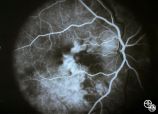 |
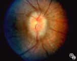
Pseudopapilledema With Retinal Neovascularization, Fluorescein Angiogram | Joel M. Weinstein, MD | This 8-year-old boy presented with a 2-week history of decreased vision in the right eye. He had undergone a normal MRI and CSF examination, including intracranial pressure, before neuro-ophthalmologic assessment. The fundus photographs and fluorescein angiograms show subretinal neovascularization a... |
| 303 |
 |
Isolated Optic Neuritis/Neuropathy | Anthony C. Arnold, MD | This 42-year-old male with pseudotumor cerebri and chronic papilledema demonstrated refractile bodies, which can be seen with chronic optic disc edema. This image shows the chronic papilledema at presentation, with associated refractile hyaline bodies at the disc periphery in both eyes. Pair with 96... |
| 304 |
 |
Isolated Congenital Optic Disc Anomalies | Joel M. Weinstein, MD | This 8-year-old boy presented with a 2-week history of decreased vision in the right eye. He had undergone a normal MRI and CSF examination, including intracranial pressure, before neuro-ophthalmologic assessment. The fundus photographs and fluorescein angiograms show subretinal neovascularization a... |
| 305 |
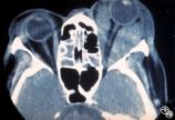 |
Ocular Manifestations of Systemic Disorders | Mitchell J. Wolin, MD | Thyroid eye disease is the most common cause of unilateral or bilateral proptosis in the adult patient. Other signs of thyroid eye disease should be sought, including lid retraction, inferior scleral show, and lid lag. Patients with markedly asymmetric or strictly unilateral proptosis should probabl... |
| 306 |
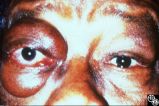 |
Ocular Manifestations of Systemic Disorders | Mitchell J. Wolin, MD | Thyroid eye disease is the most common cause of unilateral or bilateral proptosis in the adult patient. Other signs of thyroid eye disease should be sought, including lid retraction, inferior scleral show, and lid lag. Patients with markedly asymmetric or strictly unilateral proptosis should probabl... |
| 307 |
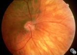 |
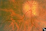
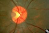
Isolated Congenital Optic Disc Anomalies | William Fletcher Hoyt, MD | This patient demonstrates bilateral tilted optic discs. Patients with this congenital optic disc anomaly may be asymptomatic or have bitemporal visual field defects that do not respect the vertical midline. Disease/Diagnosis: Tilted Discs. |
| 308 |
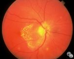 |
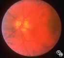
Optic Neuropathies | Ralph A. Sawyer, MD | Optic disc edema with a macular star figure has been referred to as neuroretinitis. |
| 309 |
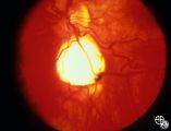 |
Retinal Coloboma Underneath a Relatively Normal Optic Nerve | Thomas R. Wolf, MD | Optic nerve colobomas appear as enlarged, white optic discs that are deeply excavated, often with some sapring of the superior rim. They result from an abnormal fusion of the proximal embryonic fissure. Optic nerve colobomas occur unilaterally or bilaterally with a similar frequency and can result i... |
| 310 |
 |
Isolated Congenital Optic Disc Anomalies | Thomas R. Wolf, MD | Patients with midline closure defects may exhibit abnormalities in the optic nerve, choroid, retinal pigment epithelium or retina. Anterior closure defects may result in colobomas of the structures of the anterior segment, such as the iris. Disease/Diagnosis: Coloboma. |
| 311 |
 |
Isolated Congenital Optic Disc Anomalies | Anthony C. Arnold, MD | A 10-year-old girl had central visual loss due to this optic pit. Disease/Diagnosis: Optic Pit. |
| 312 |
 |
Isolated Congenital Optic Disc Anomalies | Anthony C. Arnold, MD | This is a photograph of peripheral drusen. The paired image 92_69 demonstrates the typical autofluorescence. |
| 313 |
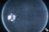 |
Optic Disc Drusen Autofluorescence | Anthony C. Arnold, MD | This case of optic disc drusen demonstrates the typical autofluorescence. Pair with 92_68. Imaging: Autoflourescence? |
| 314 |
 |
Isolated Congenital Optic Disc Anomalies | Rosa A. Tang, MD | Normally, there is no visible myelination of the nerve fiber layer in the retina. Occasionally, visible myelination occurs that takes on a characteristic white, arcuate, feathery appearance that follows the contour of the nerve fiber layer. Disease/Diagnosis: Myelinated Nerve Fiber. |
| 315 |
 |
Peripapillary Staphyloma | Thomas R. Wolf, MD | Patients with ectasia of the outer layers of the eye may exhibit a posterior protrusion that appears on funduscopy as an area of deep excavation of the retina (posterior staphyloma). When it occurs around the optic disc, as in this case, it is termed a peripapillary staphyloma. This may occur in ass... |
| 316 |
 |
Motility Disturbances | Thomas R. Wolf, MD | The patient is a 53-year-old man with diplopia from right oculomotor nerve palsy and left hemiparesis (Weber's syndrome), with associated left lung hilar mass. The spinal tap showed pleocytosis consistent with carcinomatous meningitis. This image demonstrates oculomotor nerve metastatic carcinomatos... |
| 317 |
 |
Ocular Manifestations of Congenital/Inherited Diseases | William Fletcher Hoyt, MD | Tuberous sclerosis is an autosomal dominant phakomatosis characterized by astrocytic hamartomas of the retina or optic nerve, adenoma sebaceum of the face, periungual fibromas, and astrocytic hamartomas of the brain, with secondary seizures and mental retardation. Disease/Diagnosis: Tuberous Scleros... |
| 318 |
 |
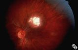
Ocular Manifestations of Congenital/Inherited Diseases | Anthony C. Arnold, MD | Note the retinal/optic nerve head astrocytoma in a 12-year-old boy with tuberous sclerosis. Disease/Diagnosis: Tuberous Sclerosis. |
| 319 |
 |
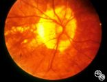
Systemic Disorders With Optic Nerve and Retinal Findings | Larry P. Frohman, MD | A 29-year-old African American woman presented with headaches, bilateral transient visual obscurations, blurred vision, numbness, and weakness of the lower extremities with myalgia and joint pains. She had an unplanned 12-pound weight loss over 2 months. A neurologist and internist diagnosed her wit... |
| 320 |
 |
Systemic Disorders With Optic Nerve and Retinal Findings | Larry P. Frohman, MD | A 29-year-old African American woman presented with headaches, bilateral transient visual obscurations, blurred vision, numbness, and weakness of the lower extremities with myalgia and joint pains. She had an unplanned 12-pound weight loss over 2 months. A neurologist and internist diagnosed her wit... |
| 321 |
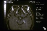 |
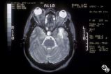
Systemic Disorders With Optic Nerve and Retinal Findings | Larry P. Frohman, MD | A 35-year-old African-American woman had gradual bilateral painless visual loss over 3 months. When initially seen, the visual acuities were HM OD, NLP OS. The MRI showed diffused enhancement of the optic nerves and lacrimal glands. The evaluation strongly suggested sarcoidosis, with elevated angiot... |
| 322 |
 |
Systemic Disorders With Optic Nerve and Retinal Findings | Larry P. Frohman, MD | A 35-year-old African-American woman had gradual bilateral painless visual loss over 3 months. When initially seen, the visual acuities were HM OD, NLP OS. The MRI showed diffused enhancement of the optic nerves and lacrimal glands. The evaluation strongly suggested sarcoidosis, with elevated angiot... |
| 323 |
 |
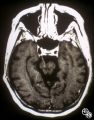
Chiasmal Syndromes | Larry P. Frohman, MD | This 36-year-old woman presented in 1988 with 3 weeks of vertical binocular diplopia. She was a known amblyope OD. Her examination was notable for a right hyperdeviation of 1 PD present in right gaze and a subtle left noncongruous homonymous field defect. She was lost to follow-up, but 5 years later... |
| 324 |
 |
Chiasmal Syndromes | Larry P. Frohman, MD | This 36-year-old woman presented in 1988 with 3 weeks of vertical binocular diplopia. She was a known amblyope OD. Her examination was notable for a right hyperdeviation of 1 PD present in right gaze and a subtle left noncongruous homonymous field defect. She was lost to follow-up, but 5 years later... |
| 325 |
 |
Systemic Disorders With Optic Nerve and Retinal Findings | Larry P. Frohman, MD | Skin rashes occur in about 30 percent of patients with sarcoid. When seen, the rashes offer an accessible site for obtaining histologic material for confirmation of the clinical diagnosis. Pair with 91_68. |