Best known for his world-renowned neuro-ophthalmology unit based at the University of California, San Francisco, William Hoyt, MD collected here more than 850 of his best images covering a wide range of disorders.
William F. Hoyt, MD, Professor Emeritus of Ophthalmology, Neurology and Neurosurgery, Department of Ophthalmology, University of California, San Francisco.
NOVEL: https://novel.utah.edu/
TO
| Title | Description | Type | ||
|---|---|---|---|---|
| 276 |
 |
Multiple Sclerosis Slits and Thinning in Peripapillary (Retinal) Nerve Riber Layer | Need magnification - Left eye - Inferior arcuate nerve fiber slits. Pairs with IIB2_01a & IIB2_01c. Anatomy: Peripapillary nerve fiber layer. Pathology: Slit-like atrophy. Disease/Diagnosis: Multiple sclerosis optic neuropathy. Clinical: No symptoms. | Image |
| 277 |
 |
Ocular Hypertension | There is an argument about whether this is a pseudo nerve fiber layer defect, because defect does not reach the disc. The vessels transversing the defect are not exposed and there is no change in the visual field and no other slit like defects. Anatomy: Peripapillary nerve fiber layer. Pathology: Sl... | Image |
| 278 |
 |
Ocular Hypertension | Need color work to show superior slit 1972. Right eye. Ocular hypertension. No field defect recognized. Anatomy: Peripapillary nerve fiber layer. Pathology: Slit-like defects in the arcuate nerve fiber bundles. Disease/Diagnosis: Ocular hypertension. Clinical: Elevated intraocular pressure. | Image |
| 279 |
 |
Ocular Hypertension | Multiple slit like defects in the upper arcuate nerve bundles. 1971. Anatomy: Peripapillary nerve fiber layer. Pathology: Slit-like defects in the arcuate nerve fiber bundles. Disease/Diagnosis: Elevated intraocular pressure. Clinical: Elevated intraocular pressure. | Image |
| 280 |
 |
Ocular Hypertension | Multiple slit defects in the upper arcuate bundles. 1972. Anatomy: Peripapillary nerve fiber layer. Pathology: Slit-like defects in the arcuate nerve fiber bundles. Disease/Diagnosis: Elevated intraocular pressure. Clinical: Elevated intraocular pressure. | Image |
| 281 |
 |
P50 Chronic Papilledema with Subretinal Neo-Vascular Network | Chronic papilledema with subretinal neo-vascular network. Pseudotumor. Anatomy: Optic disc. Pathology: Papilledema. Disease/ Diagnosis: Chronic papilledema with sub-retinal neovascular network. | Image |
| 282 |
 |
P52a Asymmetric Papilledema with Choroidal Folds | Left eye. Choroidal folds with no papilledema. Asymmetric papilledema with choroidal folds. Bilateral choroidal folds from elevated intracranial pressure. 52a. Anatomy: Optic disc. Pathology: Papilledema. Disease/ Diagnosis: Asymmetric - No papilledema with choroidal folds | Image |
| 283 |
 |
Resolution of Papilledema Following Optic Nerve Sheath Decompression (ONSD) | Left eye. 17 year old boy. Cryptococcal meningitis. Module developed papilledema. June 1974. Anatomy: Optic disc. Pathology: Papilledema. Disease/Diagnosis: Resolving papilledema. | Image |
| 284 |
 |
Resolution of Papilledema Following Optic Nerve Sheath Decompression (ONSD) | Left eye. 17 year old boy. Cryptococcal meningitis. Resolution of papilledema following optic nerve sheath fenestration (ONSF) on November 1, 1974. Same eye as P_53a on November 7, 1974, one week following ONSF. Anatomy: Optic disc. Pathology: Papilledema. Disease/Diagnosis: Resolving papilledema. | Image |
| 285 |
 |
Resolution of Papilledema Following Optic Nerve Sheath Decompression (ONSD) | Left eye. 17 year old boy. Cryptococcal meningitis. Same eye as P_53a. Increased papilledema. August 1974. Anatomy: Optic disc. Pathology: Papilledema. Disease/Diagnosis: Resolving papilledema. | Image |
| 286 |
 |
Unilateral Papilledema | Unilateral papilledema in Pseudotumor cerebri in patient with elevated intracranial pressure. Left eye. Has no optic cup. Disc is flat. Anatomy: Optic disc. Pathology: Unliateral papilledema. Disease/Diagnosis: Idiopathic intracranial hypertension (pseudotumor cerbri). Clinical: Transient monocular ... | Image |
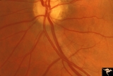
| 287 |
 |
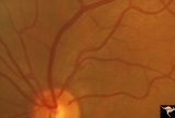
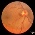
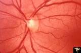
Vascular Disc Anomalies - Prepapillary Arterial Loop | Multiple prepapillary arterial loops involving superior and inferior retinal arterioles. Anatomy: Optic disc. Pathology: Congenital prepapillary arterial loop. Disease/Diagnosis: Congenital prepapillary arterial loop. Clinical: Asymptomatic. Patient presented with migraine. | Image |
| 288 |
 |
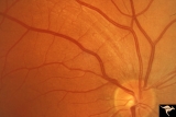
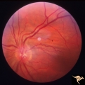
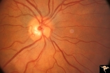
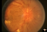
Vascular Disc Anomalies - Prepapillary Arterial Loop | Prepapillary arterial loop arising typically from the inferior retinal arterioles and projecting forward into the vitreous. 42 year old patient. Anatomy: Optic disc. Pathology: Congenital prepapillary arterial loop. Disease/Diagnosis: Congenital prepapillary arterial loop. Clinical: Asymptomatic. | Image |
| 289 |
 |
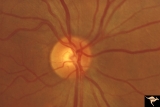
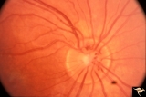
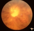
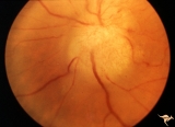
Vascular Disc Anomalies - Prepapillary Arterial Loop | Small central prepapillary arterial loop. 30 year old woman. Anatomy: Optic disc. Pathology: Congenital prepapillary arterial loop. Disease/Diagnosis: Congenital prepapillary arterial loop. Clinical: Asymptomatic. | Image |
| 290 |
 |
F102 Myeloblastic Leukemia | Myeloblastic leukemia. Left eye. Pair with F1_03. Anatomy: Optic disc. Pathology: Neoplastic (metastatic) papillopathy. Disease/ Diagnosis: Myeloblastic leukemia. | Image |
| 291 |
 |
F103 Myeloblastic Leukemia | Myeloblastic leukemia. Right eye. Pair with F1_02. Anatomy: Optic disc. Pathology: Neoplastic (metastatic) papillopathy. Disease/ Diagnosis: Myeloblastic leukemia. | Image |
| 292 |
 |
F2b13 Progression of Optic Disc Changes Caused by Malignant Optic Nerve Glioma of Adulthood | Progression. Group with F2b_12_1 and F2b_14_3. 69 year old male. April 22, 1992. There are signs of CVRO. Reference: Hoyt WF, Meshel LG, Lessell S, Schatz NJ, Suckling RD. Malignant optic glioma of adulthood. Brain. 1973;96(1):121-32. Anatomy: Optic disc. Pathology: Optic nerve glioma. Disease/ Diag... | Image |
| 293 |
 |
F2b14 Progression of Optic Disc Changes Caused by Malignant Optic Nerve Glioma of Adulthood | Progression. Group with F2b_12_1 and F2b_13_2. 69 year old male. Shows signs of myelin being squeezed through the optic disc into the eye. June 6, 1992. Reference: Hoyt WF, Meshel LG, Lessell S, Schatz NJ, Suckling RD. Malignant optic glioma of adulthood. Brain. 1973;96(1):121-32. Anatomy: Optic di... | Image |
| 294 |
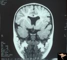 |
H07 Panhypoplasia | MRI Scan, coronal view showing absence of septum pellucidum. Hypoplastic chiasm. De Morsier's syndrome. Same patient as H_6. Anatomy: Optic disc. Pathology: Hypoplasia of the optic nerve. Disease/ Diagnosis: Hypoplasia. Imaging: MRI scan. | Image |
| 295 |
 |
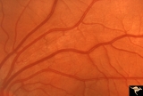
Vascular Disc Anomalies - Prepapillary Arterial Loop | Prepapillary arterial loops. 36 year old woman. Anatomy: Optic disc. Pathology: Congenital prepapillary arterial loop. Disease/Diagnosis: Congenital prepapillary arterial loop. Clinical: Asymptomatic. | Image |
| 296 |
 |
G103 Evulsion | Partial evulsion of the right optic nerve. Notice what is left of superior optic nerve. Anatomy: Optic disc. Pathology: Optic disc has been evulsed. Disease/ Diagnosis: Evulsion of the optic disc. | Image |
| 297 |
 |
Ocular Hypertension | Chronic simple glaucoma. 1976. Magnified of IIB3_3a. Note slits in upper arcuate nerve fiber layer. Pair with IIB3_3a. Anatomy: Peripapillary nerve fiber layer. Pathology: Slit-like defects in the arcuate nerve fiber bundles. Disease/Diagnosis: Elevated intraocular pressure. Clinical: Elevated intra... | Image |
| 298 |
 |
Ocular Hypertension | Chronic simple glaucoma. 1976. Note slits in upper arcuate nerve fiber layer. Pair with IIB3_3b. Anatomy: Peripapillary nerve fiber layer. Pathology: Slit-like defects in the arcuate nerve fiber bundles. Disease/Diagnosis: Elevated intraocular pressure. Clinical: Elevated intraocular pressure. | Image |
| 299 |
 |
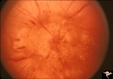
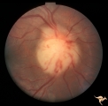
F205 Optic Nerve Sheath Meningioma | Optic nerve meningioma of right optic nerve. Progressive visual field loss. Notice macular star and "cotton wool" spots. Anatomy: Optic disc. Pathology: Chronic optic disc swelling caused by optic nerve sheath meningioma. Disease/ Diagnosis: Chronic optic disc swelling caused by optic nerve sheath m... | Image |
| 300 |
 |
F107 Metastatic Breast Cancer to the Disc | Metastatic breast cancer to the disc. Notice mass on inferior portion of disc. Also notice tangled capillary dilation within the mass indicating infiltration. This disc tumor was radiated. It disappeared leaving a pale flat atrophic nerve. The patient died. Histologic study of the eye revealed metas... | Image |