Best known for his world-renowned neuro-ophthalmology unit based at the University of California, San Francisco, William Hoyt, MD collected here more than 850 of his best images covering a wide range of disorders.
William F. Hoyt, MD, Professor Emeritus of Ophthalmology, Neurology and Neurosurgery, Department of Ophthalmology, University of California, San Francisco.
NOVEL: https://novel.utah.edu/
TO
| Title | Description | Type | ||
|---|---|---|---|---|
| 276 |
 |
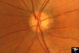
H84 Chiasmal Hemioptic Hypoplasia | Congenital bitemporal hemianopia with marked bi-nasal hypoplasia. Left eye. 17 year old male. Same patient as H_85. Anatomy: Optic disc. Pathology: Chiasmal hemioptic hypoplasia. Disease/ Diagnosis: Congenital anomaly involving chiasm. | Image |
| 277 |
 |
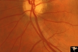
H88 Chiasmal Hemioptic Hypoplasia | Nasal hypoplasia with temporal hemianopia from a congenital Rathke Pouch Cyst. Anatomy: Optic disc. Pathology: Chiasmal hemioptic hypoplasia. Disease/ Diagnosis: Congenital anomaly involving chiasm. | Image |
| 278 |
 |
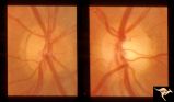
H89 Occipital Hemianoptic Hypoplasia | Diagram of homonymous hemioptic hypoplasia showing pattern of preserved nerve fibers. Homonymous hemioptic hypoplasia. Fundoscopic features in standard and red-free illumination in three patients with congenital hemiplegia. Anatomy: Optic disc. Pathology: Occipital hemianoptic hypoplasia. Disease/ D... | Image |
| 279 |
 |
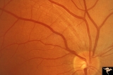
H90 Occipital Hemianoptic Hypoplasia | Note left disc (right side of image) is the eye with temporal field defect. Shows band atrophy. Anatomy: Optic disc. Pathology: Occipital hemianoptic hypoplasia. Congenital defect of the occipital lobe. | Image |
| 280 |
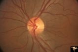 |
H91 Occipital Hemianoptic Hypoplasia | Left eye with temporal field defect shows trans-synaptic band atrophy. Same patient as H_92. Anatomy: Optic disc. Pathology: Occipital hemianoptic hypoplasia. Disease/ Diagnosis: Congenital defect of the occipital lobe. | Image |
| 281 |
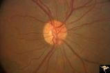 |
H92 Occipital Hemianoptic Hypoplasia | Right eye. Same patient as H_91. Anatomy: Optic disc. Pathology: Occipital hemianoptic hypoplasia. Disease/ Diagnosis: Congenital defect of the occipital lobe. | Image |
| 282 |
 |
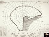
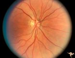
H93 Occipital Hemianoptic Hypoplasia | Visual field. Left eye. Right inferior homonymous. Same patient as H_94, H_95, H_96, H_97. Anatomy: Optic disc. Pathology: Occipital hemianoptic hypoplasia. Disease/ Diagnosis: Congenital defect of the occipital lobe. Imaging: MRI scan - See slide H97. | Image |
| 283 |
 |
H94 Occipital Hemianoptic Hypoplasia | Visual field. Right eye. Quatrantanopia. Same patient as H_93, H_95, H_96, H_97. Anatomy: Optic disc. Pathology: Occipital hemianoptic hypoplasia. Disease/ Diagnosis: Congenital defect of the occipital lobe. Imaging: MRI scan - See slide H97. | Image |
| 284 |
 |
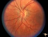
H95 Occipital Hemianoptic Hypoplasia | Right eye with temporal field defect shows trans-synaptic band atrophy. Absence of nasal nerve fibers. Same patient as H_93, H_94, H_96, H_97. Anatomy: Optic disc. Pathology: Occipital hemianoptic hypoplasia. Disease/ Diagnosis: Congenital defect of the occipital lobe. Imaging: MRI scan - See slide ... | Image |
| 285 |
 |
H96 Occipital Hemianoptic Hypoplasia | Left eye has nasal quadrantic field defect. Same patient as H_93, H_94, H_95, H_97. Anatomy: Optic disc. Pathology: Occipital hemianoptic hypoplasia. Disease/ Diagnosis: Congenital defect of the occipital lobe. Imaging: MRI scan - See slide H97. | Image |
| 286 |
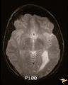 |
H97 Occipital Hemianoptic Hypoplasia | MRI scan shows left occipital lobe periventricular leuko-melacia. Same patient as H_93, H_94, H_95, H_96. Anatomy: Optic disc. Pathology: Occipital hemianoptic hypoplasia. DIsease/ Diagnosis: Congenital defect of the occipital lobe. Imaging: MRI scan. | Image |
| 287 |
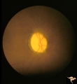 |
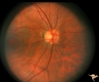
IC105 Quinine Toxicity (Amblyopia) | Quinine toxicity (amblyopia), 1971, blind eye, note diffuse arteriole narrowing and optic nerve pallor. Anatomy: Optic disc. Pathology: Toxic ischemic retinal damage. Disease/ Diagnosis: Quinine toxicity. Clinical: Blindness. | Image |
| 288 |
 |
Multiple Sclerosis Slits and Thinning in Peripapillary (Retinal) Nerve Riber Layer | Multiple slit defect in the superior arcuate nerve fiber layer. Anatomy: Peripapillary nerve fiber layer. Pathology: Slit-like atrophy. Disease/Diagnosis: Multiple sclerosis optic neuropathy. Clinical: No symptoms. | Image |
| 289 |
 |
Multiple Sclerosis Slits and Thinning in Peripapillary (Retinal) Nerve Riber Layer | Multiple slit defect in the superior arcuate nerve fiber layer. Pair with IIB2_6b. Anatomy: Peripapillary nerve fiber layer. Pathology: Slit-like atrophy. Disease/Diagnosis: Multiple sclerosis optic neuropathy. Clinical: No symptoms. | Image |
| 290 |
 |
Multiple Sclerosis Slits and Thinning in Peripapillary (Retinal) Nerve Riber Layer | Multiple slit like defects in the inferior arcuate nerve fibers. Pair with IIB2_3b. Anatomy: Peripapillary nerve fiber layer. Pathology: Slit-like atrophy. Disease/Diagnosis: Multiple sclerosis optic neuropathy. Clinical: No symptoms. | Image |
| 291 |
 |
Multiple Sclerosis Slits and Thinning in Peripapillary (Retinal) Nerve Riber Layer | Multiple slit and wedge defects in the nerve fiber layer. Pair with IIB2_3a. Anatomy: Peripapillary nerve fiber layer. Pathology: Slit-like atrophy. Disease/Diagnosis: Multiple sclerosis optic neuropathy. Clinical: No symptoms. | Image |
| 292 |
 |
Multiple Sclerosis Slits and Thinning in Peripapillary (Retinal) Nerve Riber Layer | Multiple slit defect in the superior arcuate nerve fiber layer. Anatomy: Peripapillary nerve fiber layer. Pathology: Slit-like atrophy. Disease/Diagnosis: Multiple sclerosis optic neuropathy. Clinical: No symptoms. | Image |
| 293 |
 |
Multiple Sclerosis Slits and Thinning in Peripapillary (Retinal) Nerve Riber Layer | Multiple slit defect in the superior arcuate nerve fiber layer. Magnified. Pair with IIB2_6a. Anatomy: Peripapillary nerve fiber layer. Pathology: Slit-like atrophy. Disease/Diagnosis: Sclerosis optic neuropathy. Clinical: No symptoms. | Image |
| 294 |
 |
Multiple Sclerosis Slits and Thinning in Peripapillary (Retinal) Nerve Riber Layer | Left eye. Upper arcuate nerve fiber layer contains multiple low density slits. These indicate nerve fiber loss. Anatomy: Peripapillary nerve fiber layer. Pathology: Slit-like atrophy. Disease/Diagnosis: Multiple sclerosis optic neuropathy. Clinical: No symptoms. | Image |
| 295 |
 |
Multiple Sclerosis Slits and Thinning in Peripapillary (Retinal) Nerve Riber Layer | Multiple slit defect in the superior arcuate nerve fiber layer in a 13 year old boy. Right eye. Pair with IIB2_7a. Anatomy: Peripapillary nerve fiber layer. Pathology: Slit-like atrophy. Disease/Diagnosis: Multiple sclerosis optic neuropathy. Clinical: No symptoms. | Image |
| 296 |
 |
Multiple Sclerosis Slits and Thinning in Peripapillary (Retinal) Nerve Riber Layer | Multiple slit defect in the superior arcuate nerve fiber layer in a 13 year old boy. Pair with IIB2_7b. Anatomy: Peripapillary nerve fiber layer. Pathology: Slit-like atrophy. Disease/Diagnosis: Multiple sclerosis optic neuropathy. Clinical: No symptoms. | Image |
| 297 |
 |
Multiple Sclerosis Slits and Thinning in Peripapillary (Retinal) Nerve Riber Layer | Need magnification - Left eye - Peculiar punctate dotted surface of internal limiting membrane reflexes. Pairs with IIB2_01a & IIB2_02b. Anatomy: Peripapillary nerve fiber layer. Pathology: Slit-like atrophy. Disease/Diagnosis: Multiple sclerosis optic neuropathy. Clinical: No symptoms. | Image |
| 298 |
 |
Multiple Sclerosis Slits and Thinning in Peripapillary (Retinal) Nerve Riber Layer | Need magnification - Left eye - Inferior arcuate nerve fiber slits. Pairs with IIB2_01b & IIB2_01c. Anatomy: Peripapillary nerve fiber layer. Pathology: Slit-like atrophy. Disease/Diagnosis: Multiple sclerosis optic neuropathy. Clinical: No symptoms. | Image |
| 299 |
 |
Multiple Sclerosis Slits and Thinning in Peripapillary (Retinal) Nerve Riber Layer | Need magnification - Left eye - Inferior arcuate nerve fiber slits. Pairs with IIB2_01a & IIB2_01c. Anatomy: Peripapillary nerve fiber layer. Pathology: Slit-like atrophy. Disease/Diagnosis: Multiple sclerosis optic neuropathy. Clinical: No symptoms. | Image |
| 300 |
 |
Ocular Hypertension | There is an argument about whether this is a pseudo nerve fiber layer defect, because defect does not reach the disc. The vessels transversing the defect are not exposed and there is no change in the visual field and no other slit like defects. Anatomy: Peripapillary nerve fiber layer. Pathology: Sl... | Image |