AAO-NANOS Neuro-Ophthalmology Clinical Collection: Derived from the AAO-NANOS Clinical Neuro-Ophthalmology collection produced on CD. The images are of selected cases from the NANOS teaching slide exchange, and the CD was produced under the direction of Larry Frohman, MD and Andrew Lee, MD.
The American Academy of Ophthalmology (AAO); The North American Neuro-Ophthalmology Association (NANOS).
NOVEL: https://novel.utah.edu/
TO
Filters: Collection: "ehsl_novel_aao_nanos"
| Title | Creator | Description | ||
|---|---|---|---|---|
| 276 |
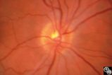 |
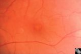
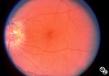
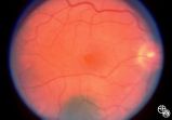
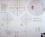
Optic Neuropathies | Daniel M. Jacobson MD | This 28-year-old otherwise-healthy woman was referred to for treatment of what was thought to be optic neuritis OD. Three weeks earlier she had noted a dark inferior scotoma OD that progressed to involve fixation over the next 10-12 days. She experienced photopsias OD at the onset. She had no viral ... |
| 277 |
 |
Optic Neuropathies | Daniel M. Jacobson MD | This 28-year-old otherwise-healthy woman was referred to for treatment of what was thought to be optic neuritis OD. Three weeks earlier she had noted a dark inferior scotoma OD that progressed to involve fixation over the next 10-12 days. She experienced photopsias OD at the onset. She had no viral ... |
| 278 |
 |
Optic Neuropathies | Daniel M. Jacobson MD | This 28-year-old otherwise-healthy woman was referred to for treatment of what was thought to be optic neuritis OD. Three weeks earlier she had noted a dark inferior scotoma OD that progressed to involve fixation over the next 10-12 days. She experienced photopsias OD at the onset. She had no viral ... |
| 279 |
 |
Optic Neuropathies | Daniel M. Jacobson MD | This 28-year-old otherwise-healthy woman was referred to for treatment of what was thought to be optic neuritis OD. Three weeks earlier she had noted a dark inferior scotoma OD that progressed to involve fixation over the next 10-12 days. She experienced photopsias OD at the onset. She had no viral ... |
| 280 |
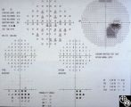 |
Optic Neuropathies | Daniel M. Jacobson MD | A previously healthy 28-year-old woman (except for a history of optic neuritis OS 9 years previously with full recovery) was referred for management of optic neuritis OD. Three weeks earlier she had noted a dark inferior scotoma OD that progressed to involve fixation over the next 10-12 days. She ex... |
| 281 |
 |
Optic Neuropathies | Daniel M. Jacobson MD | This 28-year-old otherwise-healthy woman was referred to for treatment of what was thought to be optic neuritis OD. Three weeks earlier she had noted a dark inferior scotoma OD that progressed to involve fixation over the next 10-12 days. She experienced photopsias OD at the onset. She had no viral ... |
| 282 |
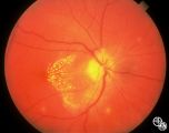 |
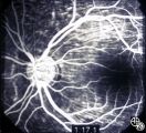
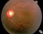
Optic Neuropathies | Ralph A. Sawyer, MD | Optic disc edema with a macular star figure has been referred to as neuroretinitis. |
| 283 |
 |
Optic Neuropathies | Michael Wall, MD | Optic disc edema with a macular star figure may occur in infectious diseases (eg, cat-scratch disease, syphilis, tuberculosis, Lyme disease), inflammatory diseases (eg, sarcoid), ischemic diseases (anterior ischemic optic neuropathy), and in papilledema. Infectious causes should be sought in patient... |
| 284 |
 |
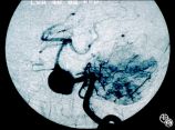
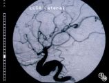
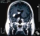
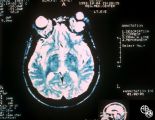
Optic Tract Syndrome Due to Carotid Artery Dolichoectasia | Larry P. Frohman, MD | This 43-year-old man was referred for evaluation of 6 months of visual loss OU. In retrospect, he had noticed increasing difficulty with his tennis game dating back over 3 years, as balls would pass him unexpectedly when hit to his backhand (left) side. The patient did not think this was progressive... |
| 285 |
 |
Optic Tract Syndrome Due to Carotid Artery Dolichoectasia | Larry P. Frohman, MD | This 43-year-old man was referred for evaluation of 6 months of visual loss OU. In retrospect, he had noticed increasing difficulty with his tennis game dating back over 3 years, as balls would pass him unexpectedly when hit to his backhand (left) side. The patient did not think this was progressive... |
| 286 |
 |
Optic Tract Syndrome Due to Carotid Artery Dolichoectasia | Larry P. Frohman, MD | This 43-year-old man was referred for evaluation of 6 months of visual loss OU. In retrospect, he had noticed increasing difficulty with his tennis game dating back over 3 years, as balls would pass him unexpectedly when hit to his backhand (left) side. The patient did not think this was progressive... |
| 287 |
 |
Optic Tract Syndrome Due to Carotid Artery Dolichoectasia | Larry P. Frohman, MD | This 43-year-old man was referred for evaluation of 6 months of visual loss OU. In retrospect, he had noticed increasing difficulty with his tennis game dating back over 3 years, as balls would pass him unexpectedly when hit to his backhand (left) side. The patient did not think this was progressive... |
| 288 |
 |
Optic Tract Syndrome Due to Carotid Artery Dolichoectasia | Larry P. Frohman, MD | This 43-year-old man was referred for evaluation of 6 months of visual loss OU. In retrospect, he had noticed increasing difficulty with his tennis game dating back over 3 years, as balls would pass him unexpectedly when hit to his backhand (left) side. The patient did not think this was progressive... |
| 289 |
 |
Optic Tract Syndrome Due to Carotid Artery Dolichoectasia | Larry P. Frohman, MD | This 43-year-old man was referred for evaluation of 6 months of visual loss OU. In retrospect, he had noticed increasing difficulty with his tennis game dating back over 3 years, as balls would pass him unexpectedly when hit to his backhand (left) side. The patient did not think this was progressive... |
| 290 |
 |
Optic Tract Syndrome Due to Carotid Artery Dolichoectasia | Larry P. Frohman, MD | This 43-year-old man was referred for evaluation of 6 months of visual loss OU. In retrospect, he had noticed increasing difficulty with his tennis game dating back over 3 years, as balls would pass him unexpectedly when hit to his backhand (left) side. The patient did not think this was progressive... |
| 291 |
 |
Optic Tract Syndrome Due to Carotid Artery Dolichoectasia | Larry P. Frohman, MD | This 43-year-old man was referred for evaluation of 6 months of visual loss OU. In retrospect, he had noticed increasing difficulty with his tennis game dating back over 3 years, as balls would pass him unexpectedly when hit to his backhand (left) side. The patient did not think this was progressive... |
| 292 |
 |
Orbital Tumors | Mitchell J. Wolin, MD | Cavernous hemangiomas of the orbit usually result in painless orbital signs such as proptosis or visual loss. Orbital imaging of the lesion, which usually is a well-defined orbital mass, is demonstrated in this study. The lesion is benign and usually occurs in young to middle-aged adults. Surgical e... |
| 293 |
 |
Orbital Tumors | Mitchell J. Wolin, MD | Cavernous hemangiomas of the orbit usually result in painless orbital signs such as proptosis or visual loss. Orbital imaging of the lesion, which usually is a well-defined orbital mass, is demonstrated in this study. The lesion is benign and usually occurs in young to middle-aged adults. Surgical e... |
| 294 |
 |
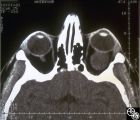
Orbital Tumors | Larry P. Frohman, MD | This 30-year-old man had a retrobulbar intraconal mass OS. The CT scans showed a heterogeneous lobulated enhancing mass, 2.2 x 1.9 x 1.8 cm. The case beautifully exhibits chorodial folds. The ultrasound showed internal reflectivity. The patient refused surgery. Pair with Images 97_60, 97_62, 97_63, ... |
| 295 |
 |
Orbital Tumors | Larry P. Frohman, MD | This 30-year-old man had a retrobulbar intraconal mass OS. The CT scans showed a heterogeneous lobulated enhancing mass, 2.2 x 1.9 x 1.8 cm. The case beautifully exhibits chorodial folds. The ultrasound showed internal reflectivity. The patient refused surgery. Pair with Images 97_60, 97_61, 97_63, ... |
| 296 |
 |
Orbital Tumors | Larry P. Frohman, MD | This 30-year-old man had a retrobulbar intraconal mass OS. The CT scans showed a heterogeneous lobulated enhancing mass, 2.2 x 1.9 x 1.8 cm. The case beautifully exhibits chorodial folds. The ultrasound showed internal reflectivity. The patient refused surgery. Pair with Images 97_61, 97_62, 97_63, ... |
| 297 |
 |
Orbital Tumors | Mitchell J. Wolin, MD | Cavernous hemangiomas of the orbit usually result in painless orbital signs such as proptosis or visual loss. Orbital imaging of the lesion, which usually is a well-defined orbital mass, is demonstrated in this study. The lesion is benign and usually occurs in young to middle-aged adults. Surgical e... |
| 298 |
 |
Orbital Tumors | Larry P. Frohman, MD | This 30-year-old man had a retrobulbar intraconal mass OS. The CT scans showed a heterogeneous lobulated enhancing mass, 2.2 x 1.9 x 1.8 cm. The case beautifully exhibits chorodial folds. The ultrasound showed internal reflectivity. The patient refused surgery. Pair with Images 97_60, 97_61, 97_62, ... |
| 299 |
 |
Orbital Tumors | Larry P. Frohman, MD | This 30-year-old man had a retrobulbar intraconal mass OS. The CT scans showed a heterogeneous lobulated enhancing mass, 2.2 x 1.9 x 1.8 cm. The case beautifully exhibits chorodial folds. The ultrasound showed internal reflectivity. The patient refused surgery. Pair with Images 97_60, 97_61, 97_62, ... |
| 300 |
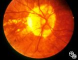 |
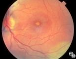
Peripapillary Staphyloma | Thomas R. Wolf, MD | Patients with ectasia of the outer layers of the eye may exhibit a posterior protrusion that appears on funduscopy as an area of deep excavation of the retina (posterior staphyloma). When it occurs around the optic disc, as in this case, it is termed a peripapillary staphyloma. This may occur in ass... |