A collection of videos relating to the diagnosis and treatment of eye movement disorders. This collection includes many demonstrations of examination techniques.
Dan Gold, D.O., Associate Professor of Neurology, Ophthalmology, Neurosurgery, Otolaryngology - Head & Neck Surgery, Emergency Medicine, and Medicine, The Johns Hopkins School of Medicine.
A collection of videos relating to the diagnosis and treatment of eye movement disorders.
NOVEL: https://novel.utah.edu/
TO
| Title | Description | Type | ||
|---|---|---|---|---|
| 276 |
 |
Oculopalatal Tremor with Prominent Nystagmus, Bilateral Horizontal Gaze Palsy, and Bilateral Facial Palsies | 𝗢𝗿𝗶𝗴𝗶𝗻𝗮𝗹 𝗗𝗲𝘀𝗰𝗿𝗶𝗽𝘁𝗶𝗼𝗻: This is a 50-year-old woman who experienced the acute onset of right sixth and seventh nerve palsies and left hemiparesis. Two cavernomas within the right pons (one in the region of the facial colliculus) were demonstrat... | Image/MovingImage |
| 277 |
 |
Oculopalatal Tremor and Internuclear Ophthalmoplegia Due to Hemorrhagic Pontine Cavernoma | 𝗢𝗿𝗶𝗴𝗶𝗻𝗮𝗹 𝗗𝗲𝘀𝗰𝗿𝗶𝗽𝘁𝗶𝗼𝗻: This is a 60-year-old woman who experienced 2 episodes of vertigo, nausea and vomiting, which was felt to be related to recurrent hemorrhage of a pontine cavernoma that was adjacent to the fourth ventricle. The cavernoma ... | Image/MovingImage |
| 278 |
 |
Eye Closure and Oculopalatal Tremor | 𝗢𝗿𝗶𝗴𝗶𝗻𝗮𝗹 𝗗𝗲𝘀𝗰𝗿𝗶𝗽𝘁𝗶𝗼𝗻: This patient suffered a traumatic brain injury with brainstem injury resulting in damage to Mollaret's triangle and palatal tremor. Inferior olivary hypertrophy was noted on her MRI, although no vertical and/or torsional ... | Image/MovingImage |
| 279 |
 |
Pontine Hemorrhage Causing Oculopalatal Tremor and Multiple Cranial Neuropathies | This is a 45-yo-woman who had a dorsal pontine cavernoma that bled 2 years prior to this video. Symptoms included diplopia and oscillopsia. On examination, she had left>right facial palsies (upper and lower face from involvement of the nucleus/fascicle - i.e., lower motor neuron palsies) and sixth n... | Image/MovingImage |
| 280 |
 |
Palato-ocular Synchrony in Oculopalatal Tremor | This is a patient with OPT due to a pontine hemorrhage, and although she did have torsional pendular nystagmus, it was very subtle. However, with eyelid closure, much larger vertical ocular oscillations could be seen, which were in fact synchronous with her palatal tremor. This finding, sometimes ... | Image/MovingImage |
| 281 |
 |
Vertical Gaze Palsy and Saccadic Intrusions Due to Anti-Ri from Head and Neck Carcinoma | A 55-yo- woman was admitted for imbalance and double vision. Three weeks prior to presentation she first noticed swelling on the right side of her face and neck. CT of the head and neck showed right-sided cervical adenopathy and enlarged left retropharyngeal node. Ultrasound- guided biopsy of the n... | Image/MovingImage |
| 282 |
 |
Upbeat Nystagmus & Ocular Flutter Due to Cerebellar Pilocytic Astrocytoma | This is a 20-year-old woman who was diagnosed with a cerebellar pilocytic astrocytoma at age 10 after presenting with severe headaches and hydrocephalus. She underwent incomplete resection and radiation therapy at that time. She experienced mild vertical oscillopsia in upgaze at baseline, and increa... | Image/MovingImage |
| 283 |
 |
Post-infectious Ocular Flutter and Myoclonus Syndrome | 𝗢𝗿𝗶𝗴𝗶𝗻𝗮𝗹 𝗗𝗲𝘀𝗰𝗿𝗶𝗽𝘁𝗶𝗼𝗻: This is a 35-yo-woman presenting with oscillopsia following a viral illness. She described being easily startled, with "shakiness" of the head/neck and body. She had myoclonus and ocular flutter, with the latter evident w... | Image/MovingImage |
| 284 |
 |
Convergence Insufficiency and Square Wave Jerks in PSP | 𝗢𝗿𝗶𝗴𝗶𝗻𝗮𝗹 𝗗𝗲𝘀𝗰𝗿𝗶𝗽𝘁𝗶𝗼𝗻: This is a 70-yo-woman with progressive supranuclear palsy with complaints of difficulty reading. Her husband noticed that she would frequently close one eye when attempting to read, and words were not clear on the page, a... | Image/MovingImage |
| 285 |
 |
Ocular Motor Signs in Progressive Supranuclear Palsy (PSP) | 𝗢𝗿𝗶𝗴𝗶𝗻𝗮𝗹 𝗗𝗲𝘀𝗰𝗿𝗶𝗽𝘁𝗶𝗼𝗻: This is a 65-yo-woman complaining of imbalance and double vision. She had significant convergence insufficiency (and would close her right eye with near viewing), providing an explanation for her diplopia. Convergence ins... | Image/MovingImage |
| 286 |
 |
Saccadic Intrusions with an Intersaccadic Interval | 𝗢𝗿𝗶𝗴𝗶𝗻𝗮𝗹 𝗗𝗲𝘀𝗰𝗿𝗶𝗽𝘁𝗶𝗼𝗻: Seen here are patients with saccadic intrusions that have preserved intersaccadic intervals. Although square wave jerks (SWJ) are present in everyone to some degree at times, when prominent or when they interfere with vis... | Image/MovingImage |
| 287 |
 |
Examples of Patients with Saccadic Intrusions (Square Wave Jerks) | Seen here are patients with saccadic intrusions that do have an intersaccadic interval. Square wave jerks are commonly seen in degenerative conditions, mainly involving the posterior fossa (e.g., cerebellar degeneration) and basal ganglia (e.g., progressive supranuclear palsy). | Image/MovingImage |
| 288 |
 |
Opsoclonus Provoked by Convergence | 𝗢𝗿𝗶𝗴𝗶𝗻𝗮𝗹 𝗗𝗲𝘀𝗰𝗿𝗶𝗽𝘁𝗶𝗼𝗻: This is a 40-yo-man with post-infectious opsoclonus-myoclonus syndrome. Opsoclonus was intermittently evident in primary position, but was consistently provoked (and intensified) by convergence. Occasionally, opsoclonus (... | Image/MovingImage |
| 289 |
 |
Chronic Progressive External Ophthalmoplegia (CPEO) and Cerebellar Signs | This is a 60-yo-woman who initially presented with imbalance and ophthalmoparesis. Initially, there was mild horizontal gaze limitation with mild gaze-evoked nystagmus and slow saccades, and over the years, gait ataxia and dysarthria (mainly a scanning quality to her speech) developed, and her ophth... | Image/MovingImage |
| 290 |
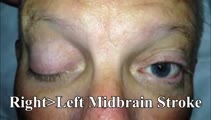 |
Bilateral Pseudo-abducens Palsies Due to Midbrain Stroke | 𝗢𝗿𝗶𝗴𝗶𝗻𝗮𝗹 𝗗𝗲𝘀𝗰𝗿𝗶𝗽𝘁𝗶𝗼𝗻: This is a man who suffered right>left midbrain strokes due to endocarditis complaining of ptosis and inability to move his eyes as well as hallucinations (peduncular hallucinosis). There was a presumed nuclear 3rd nerve p... | Image/MovingImage |
| 291 |
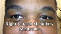 |
Saccadic Dysmetria and Ocular Lateropulsion in Lateral Medullary Stroke | 𝗢𝗿𝗶𝗴𝗶𝗻𝗮𝗹 𝗗𝗲𝘀𝗰𝗿𝗶𝗽𝘁𝗶𝗼𝗻: This is a 30-yo-man who suffered a right lateral medullary stroke. Examination showed saccadic hypermetria to the right (ipsilesional), hypometria to the left (contralesional)and rightward ocular lateropulsion (ipsilesion... | Image/MovingImage |
| 292 |
 |
Slow Horizontal, Vertical, Oblique Saccades and Gaze-evoked Nystagmus in Anti-AGNA-1 Encephalitis | This is a patient who presented subacutely with imbalance and dizziness. On examination, she had evidence of gaze evoked nystagmus, right internuclear ophthalmoplegia, as well as slow saccades horizontally and vertically. She was diagnosed with a rare antibody-mediated disorder, anti-AGNA-1 (antig... | Image/MovingImage |
| 293 |
 |
Neuro-Ophthalmic Features and Pseudo-MG Lid Signs in Miller Fisher Syndrome | 𝗢𝗿𝗶𝗴𝗶𝗻𝗮𝗹 𝗗𝗲𝘀𝗰𝗿𝗶𝗽𝘁𝗶𝗼𝗻: This is a 51-year-old woman who presented with imbalance, acute onset dizziness and diplopia that developed over three days following two weeks of upper respiratory infection and bacterial conjunctivitis. When she was ini... | Image/MovingImage |
| 294 |
 |
Miller Fisher Syndrome - Ophthalmoplegia and Hyperreflexia | 𝗢𝗿𝗶𝗴𝗶𝗻𝗮𝗹 𝗗𝗲𝘀𝗰𝗿𝗶𝗽𝘁𝗶𝗼𝗻: This is a 45-yo-woman who presented with mild imbalance and diplopia. There had been a preceding viral illness several weeks prior. Examination demonstrated horizontal gaze paresis (sparing unilateral adduction), mild gai... | Image/MovingImage |
| 295 |
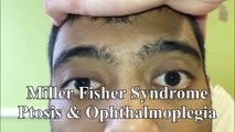 |
Miller Fisher Syndrome - Ophthalmoplegia, Ptosis and Ataxia | 𝗢𝗿𝗶𝗴𝗶𝗻𝗮𝗹 𝗗𝗲𝘀𝗰𝗿𝗶𝗽𝘁𝗶𝗼𝗻: This is a young man who presented with ptosis, difficulty moving the eyes and gait imbalance several weeks after a GI illness. Miller Fisher syndrome was diagnosed, IVIG therapy was initiated, and anti-Gq1b antibodies cam... | Image/MovingImage |
| 296 |
 |
Upbeat Transitioning to Downbeat Nystagmus in Wernicke's Encephalopathy | This is a 30-year-old man with a history of alcohol abuse who presented to the hospital with inability to walk after several weeks of heavy drinking and malnutrition. While in the hospital, he noted that when he would look straight ahead, everything he saw would appear to be bouncing up and down - a... | Image/MovingImage |
| 297 |
 |
Slow Volitional Saccades and Poor Fast Phases to an Optokinetic Stimulus, with Preserved Head Impulse Testing | This is a 67-year-old woman presenting with imbalance and binocular horizontal diplopia at near. On examination there were frequent square wave jerks, limited supraduction OU and convergence insufficiency, which explained her diplopia. Pursuit and suppression of the vestibulo-ocular reflex were sa... | Image/MovingImage |
| 298 |
 |
Trigeminal Neuropathy with Loss of the Corneal Reflex | This is a woman who underwent radiofrequency ablation for left trigeminal neuralgia. Examination demonstrated loss of facial sensation on the left in addition to an absent corneal reflex on the left, consistent with involvement of the V1 (ophthalmic) branch of the trigeminal nerve. When the cornea i... | Image/MovingImage |
| 299 |
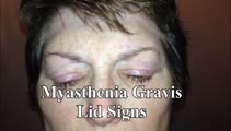 |
Typical Lid Signs (Cogan's Lid Twitch, Lid Hopping, Enhanced Ptosis) in Myasthenia Gravis | 𝗢𝗿𝗶𝗴𝗶𝗻𝗮𝗹 𝗗𝗲𝘀𝗰𝗿𝗶𝗽𝘁𝗶𝗼𝗻: This is a 60-yo-woman with MG who displays typical eyelid signs including Cogan's lid twitch, lid hopping (appreciated during horizontal smooth pursuit in this patient), and enhanced ptosis in accordance with Hering's law... | Image/MovingImage |
| 300 |
 |
Enhanced Ptosis in Myasthenia Gravis | This is a 20-yo-woman who presented with generalized weakness, ptosis and ophthalmoplegia. She had severe ptosis OU at baseline, but when one eyelid was manually elevated, there was marked enhanced ptosis of the opposite eyelid. This was in accordance with Hering's law of equal innervation of the le... | Image/MovingImage |