Best known for his world-renowned neuro-ophthalmology unit based at the University of California, San Francisco, William Hoyt, MD collected here more than 850 of his best images covering a wide range of disorders.
William F. Hoyt, MD, Professor Emeritus of Ophthalmology, Neurology and Neurosurgery, Department of Ophthalmology, University of California, San Francisco.
NOVEL: https://novel.utah.edu/
TO
Filters: Collection: "ehsl_novel_wfh"
| Title | Description | Type | ||
|---|---|---|---|---|
| 251 |
 |
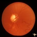
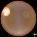
Drusen with Sub-retinal Neovascular Net | Buried drusen with sub-retinal neovascular net. There may be retinoschisis as well. Anatomy: Optic disc. Pathology: Drusen plus neovascularization at the border of the optic disc. Disease/Diagnosis: Drusen of the optic disc. Clinical: Patient has very large blind spot and impaired central vision. | Image |
| 252 |
 |
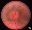
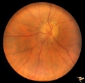
H29 Dysplasia with Hypoplasia (Elevated Dysplasia with Anomalous Vessels) | Right eye. Dysplasia with anomalous cilioretinal arterioles. Central retinal artery may be absent. Same patient as H_30. Anatomy: Optic disc. Pathology: Dysplasia of the optic disc. Disease/ Diagnosis: Elevated dysplasia with hypoplasia. | Image |
| 253 |
 |
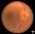
H30 Dysplasia with Hypoplasia (Elevated Dysplasia with Anomalous Vessels) | Left eye. Elevated dysplasia with anomalous cilioretinal vessels. Central retinal artery may be absent. Same patient as H_29. Anatomy: Optic disc. Pathology: Dysplasia of the optic disc. Disease/ Diagnosis: Elevated dysplasia with hypoplasia. | Image |
| 254 |
 |
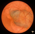
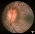
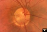
Post Papilledema with Choroidal Folds | Right eye. Post papilledema with choroidal folds due to brain tumor. Anatomy: Optic disc. Pathology: Post papilledema. Disease/Diagnosis: Post papilledema with choroidal folds. | Image |
| 255 |
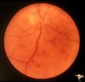 |
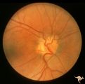
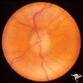
Slow Flow (Chronic Hypoxic) Retinopathy | Slow flow (chronic hypoxic) retinopathy from macroglobulanemia. Note the dot and blot hemorrhages. Anatomy: Retina. Pathology: Macroglobulanemia. Disease/Diagnosis: Slow flow (chronic hypoxic) retinopathy from macroglobulanemia. | Image |
| 256 |
 |
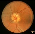
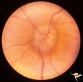
Unilateral Papilledema | Right eye. Atrophic nerve right eye. Large falx meningioma. True Foster Kennedy Syndrome. Anatomy: Optic disc. Pathology: Chronic papilledema; optic atrophy. Disease/Diagnosis: Meningioma causing Foster-Kennedy Syndrome. Clinical: Visual loss one eye; Transient visual obscuration other eye. | Image |
| 257 |
 |
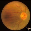
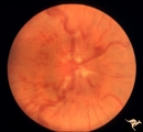
Unilateral Papilledema | Left eye. Left eye has papilledema. Large falx meningioma. True Foster Kennedy Syndrome. Anatomy: Optic disc. Pathology: Chronic papilledema; optic atrophy. Disease/Diagnosis: Meningioma causing Foster-Kennedy Syndrome. Clinical: Visual loss one eye; transient visual obscuration other eye. | Image |
| 258 |
 |
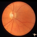
Unilateral Papilledema | Right eye. Has slight disc blur. Asymmetric papilledema. 35 year old woman. Anatomy: Optic disc. Pathology: Unilateral papilledema. Disease/Diagnosis: Idiopathic intracranial hypertension (pseudotumor cerebri). Clinical: Gaze evoked blindness. | Image |
| 259 |
 |
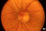
Unilateral Papilledema | Left eye. This eye has papilledema. 35 year old woman. Anatomy: Optic disc. Pathology: Unilateral papilledema. Disease/Diagnosis: Idiopathic intracranial hypertension (pseudotumor cerebri). Clinical: Gaze evoked blindness. | Image |
| 260 |
 |
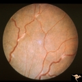
Vascular Disc Anomalies - Prepapillary Arterial Convolutions | Prepapillary arterial convolutions. Left eye. Man. Anatomy: Optic disc. Pathology: Congenital prepapillary arterial convolutions. Disease/Diagnosis: Congenital arterial vascular anomaly. Clinical: Asymptomatic. | Image |
| 261 |
 |
H25 Dysplasia with Hypoplasia (Elevated Dysplasia with Anomalous Vessels) | Right eye. Elevated dysplasia with anomalous blood vessel pattern and peri-papillary choroidal malformation. Same patient as H_26. Anatomy: Optic disc. Pathology: Dysplasia of the optic disc. Disease/ Diagnosis: Elevated dysplasia with hypoplasia. | Image |
| 262 |
 |
H26 Dysplasia with Hypoplasia (Elevated Dysplasia with Anomalous Vessels) | Left eye. Dysplasia with grossly anomalous vascular pattern. Elevated dysplasia. Same patient as H_25. Anatomy: Optic disc. Pathology: Dysplasia of the optic disc. Disease/ Diagnosis: Elevated dysplasia with hypoplasia. | Image |
| 263 |
 |
C302 Nodular Papillopathies (Sarcoid) | Perivenous Inflammatory Cuffing in a Patient with Proven Sarcoid. Left eye. Pair with C3_01. Anatomy: Retina. Pathology: Axoplasmic stasis due to sarcoid infiltration with retinal venous exudation? Disease/ Diagnosis: Sarcoid papillopathy with perivenous inflammatory disease. Clinical: Unknown? | Image |
| 264 |
 |
C301 Nodular Papillopathies (Sarcoid) | Disc swelling. Sarcoid papillopathy. Note infiltrative nodule at 9:00 on the disc.The patient had proven sarcoid. Perivenous inflammatory cuffing visible on image C3_02. Right eye. Pair with C3_02. Anatomy: Optic disc; Retina. Pathology: Axoplasmic stasis due to sarcoid infiltration. Disease/ Diagn... | Image |
| 265 |
 |
Buried Drusen | 5 year old boy. Bilateral buried drusen. Notice the lumpy nasal disc elevation. This patient had a twin brother whose optic disc drusen were exposed. Anatomy: Optic disc. Pathology: Drusen of the optic disc. Disease/Diagnosis: Drusen of the optic disc. Clinical notes: Normally functioning eye with ... | Image |
| 266 |
 |
Buried Drusen | 5 year old boy. Bilateral buried drusen. Notice the lumpy nasal disc elevation. This patient had a twin brother whose optic disc drusen were exposed. Anatomy: Optic disc. Pathology: Drusen of the optic disc. Disease/Diagnosis: Drusen of the optic disc. Clinical notes: Normally functioning eye with ... | Image |
| 267 |
 |
Chronic Papilledema with Pseudo Drusen | Left eye. Chronic papilledema with pseudo drusen due to cerebral pontine angle tumor. Anatomy: Optic disc. Pathology: Papilledema Disease/Diagnosis: Chronic papilledema with pseudo drusen. | Image |
| 268 |
 |
Chronic Papilledema with Pseudo Drusen | Right eye. Chronic papilledema with pseudo drusen due to cerebral pontine angle tumor. Anatomy: Optic disc Pathology: Papilledema Disease/Diagnosis: Chronic papilledema with pseudo drusen | Image |
| 269 |
 |
E09 Disc Swelling with Central Vein Occlusion | Chronic disc swelling due to CRVO. Anatomy: Optic disc; Retina. Pathology: Central retinal vein occlusion. Disease/ Diagnosis: Disc swelling due to central retinal vein occlusion. | Image |
| 270 |
 |
Sturge Weber Syndrome (Encephalotrigeminal Angiomatosis) | Sturge Weber Syndrome (Encephalotrigeminal angiomatosis); Color of the retina is deep red (sometimes called tomato catsup) due to a four fold thickening of the choroidal vascular bed. Glaucomatous cupping of the optic nerve. Striking retinal venous vascular anomalies on the disc and in the retina. ... | Image |
| 271 |
 |
Bilateral Papilledema | Left eye. Bilateral Papilledema with hypoparathyroidism. Woman. Anatomy: Optic disc. Pathology: Papilledema. Papilledema with hypopararthyroidism. | Image |
| 272 |
 |
Bilateral Papilledema | Right eye. Bilateral Papilledema with hypoparathyroidism. Woman. Anatomy: Optic disc. Pathology: Papilledema. Disease/Diagnosis: Papilledema with hypoparathyroidism. | Image |
| 273 |
 |
C01 Pits of the Optic Disc | Right eye. Very large inferior temporal optic pit. Congenital. Woman. Anatomy: Optic disc. | Image |
| 274 |
 |
Hereditary Macular Degenerative Disease with Spastic Paraplegia | Hereditary macular degenerative disease with Patient has spastic paraplegia associated with hereditary macular degenerative disease. Anatomy: Retina. Pathology: Cerebellar spinal degenerative disease. Disease/Diagnosis: Retinitis pigmentosa with spinal degeneration. Clinical: Hereditary spastic para... | Image |
| 275 |
 |
Ischemic Complication of Drusen | PP30a: right eye--buried drusen; PP30b: buried drusen with anterior ischemic optic neuropathy (AION) from complication of drusen of left eye. Ischemic complication of drusen in left eye. PP30c: 3 month follow-up: narrowed arterioles slightly pale disc with buried drusen. Anatomy: Optic disc. Patho... | Image |