Collection of materials relating to neuro-ophthalmology as part of the Neuro-Ophthalmology Virtual Education Library.
NOVEL: https://novel.utah.edu/
TO
- NOVEL720
| Title | Creator | Description | Subject | ||
|---|---|---|---|---|---|
| 251 |
 |
Wallenberg Syndrome and Skew Deviation | Lauren Schneider, MD; Rudrani Banik, MD | Power point of case presentation of acute Wallenberg Syndrome associated with vertical diplopia, found by 3 step and supine testing to be consistent with skew deviation. | Wallenberg Syndrome; Skew Deviation; Vertical Diplopia |
| 252 |
 |
Neuromyelitis Optica | Omar Ozgur, MD; Rudrani Banik, MD | Power point of case presentation of female with bilateral, sequential atypical optic neuritis. MRI Brain normal with no demyelination; MRI Spine shows enhancement at multiple levels and NMO antibody positive, confirming diagnosis of neuromyelitis optica (NMO). History of NMO discussed, diagnostic c... | Neuromyelitis Optica; Atypical Optic Neuritis; MRI; Plasmapheresis |
| 253 |
 |
Acute Zonal Occult Outer Retinopathy (AZOOR) versus Multiple Evanescent White Dot Syndrome (MEWDS) | Asim V. Farooq, MD; Michael T. Andreoli, MD; Heather E. Moss, MD | PPT case report on acute zonal occult outer retinopathy (AZOOR) versus multiple evanescent white dot syndrome (MEWDS). | AZOOR; MEWDS; Paracentral Scotoma; Goldmann Visual Field; Photoreceptor Loss |
| 254 |
 |
Prolactinoma in Pregnancy | Timothy Sullivan, MD; Rudrani Banik, MD | Power point of case of prolactinoma which became symptomatic during pregnancy with visual field loss. Discussion of prolactinomas and their management. Patient underwent observation only. Post-partum examination revealed resolution of bitemporal field defect with reduction in size of prolactinoma ... | Prolactinoma; Pregnancy; Bitemporal Defect |
| 255 |
 |
Cone Dystrophy | Gregory P. Van Stavern, MD | PowerPoint discussing Cone Dystrophy: Early loss of central and color vision; Color impairment often out of proportion to loss of VA; Hemeralopia ("day blindness") prominent; Light sensitivity and photophobia; Macular changes variable, and may occur late- may "Bull's Eye" pattern; Abnormal Photost... | Cone Dystrophy; Occult Macular Dystrophy; Central Cone Dystrophy |
| 256 |
 |
Pupillary Light Reflex | Wade Crow, MD | Illustration of the Pupillary Light Reflex. | Pupillary Light Reflex |
| 257 |
 |
Acute Multifocal Pigment Epithelium Epitheliopathy (AMPEE) | Gregory P. Van Stavern, MD | Images providing example of Acute Multifocal Pigment Epithelium Epitheliopathy (AMPEE) | Acute Multifocal Pigment Epithelium Epitheliopathy (AMPEE) |
| 258 |
 |
Histoplasmosis | Gregory P. Van Stavern, MD | Histoplasmosis, a fungus, can present acutely as a systemic condition. This image shows signs of Histoplasmosis. | Histoplasmosis |
| 259 |
 |
Multifocal Choroiditis | Gregory P. Van Stavern, MD | Multi-focal choroiditis is usually a bilateral choroidopathy seen more frequently in women associated with punched out appearing lesions occasionally with pigment around the edges. Image provides example. | Multi-Focal Choroiditis Panuveitis |
| 260 |
 |
Retinitis Pigmentosa | Gregory P. Van Stavern, MD | Retinitis pigmentosa is a retinal/choroidal degeneration caused by various genetic defects. The term retinitis pigmentosa is really a misnomer since it is not inflammation (retinitis) and it is not a disease of the pigmentary system (pigmentosa). | Retinitis Pigmentosa |
| 261 |
 |
Serpiginous Choroidopathy | Gregory P. Van Stavern, MD | Serpiginous choroidopathy (also known as Geographic choroidopathy) usually affects the choroid, the choriocapillaris and the retinal pigment epithelium in both eyes. | Serpiginous Choroidopathy |
| 262 |
 |
Birdshot | Gregory P. Van Stavern, MD | Birdshot Retinochoroidopathy is a posterior uveitis seen in women 30-60 years of age who present with floaters, changes in color vision, and difficulty with night vision. | Birdshot Choroidopathy |
| 263 |
 |
Pars Planitis | Gregory P. Van Stavern, MD | Pars planitis is an inflammatory condition seen in children and young adults. It is associated with inflammation of the pars plana--at the far periphery of the retina. | Pars Planitis |
| 264 |
 |
Vogt Koyanagi-Harada (VKH) Syndrome | Gregory P. Van Stavern, MD | Vogt-Koyanagi disease causes bilateral uveitis, along with alopecia, vitiligo, and hearing loss. | Vogt Koyanagi-Harada Syndrome (VKH) |
| 265 |
 |
Stargardt's Disease | Gregory P. Van Stavern, MD | Stargardt's disease is an inherited maculopathy which frequently presents with a loss of central vision. | Stargardt's Disease |
| 266 |
 |
Retinitis Pigmentosa - Rod Dystrophy | Gregory P. Van Stavern, MD | PowerPoint discussing retinitis pigmentosa, rod dystrophy. Retinitis Pigmentosa is a generalized retinal dystrophy with peripheral rather than central onset Primarily rod-cone dystrophy. Provides images. | Rod Dystrophy; Rod Dystrophy; Retinitis Pigmentosa; Night Dlindness |
| 267 |
 |
Bardet-Biedl Syndrome | Gregory P. Van Stavern, MD | PowerPoint discussing Bardet-Biedl Syndrome, a hereditary condition characterized by rod-cone dystrophy (RP), truncal obesity, polydactyly, hypogonadotropic hypogonadism (males), GU abnormalities (females), and cognitive impairment | Bardet-Biedl Syndrome; Genetics |
| 268 |
 |
Usher Syndrome | Gregory P. Van Stavern, MD | Powerpoint describing Usher Syndrome, a hereditary condition characterized by congenital, bilateral, and profound sensorineural hearing loss, adolescent onset Retinitis Pigmentosa (RP) and vestibular areflexia | Usher Syndrome; Retinal Dystrophy; Retinitis Pigmentosa; Hearing Loss |
| 269 |
 |
Vision and Alzheimer's Disease | Victoria S. Pelak, MD | Slideshow describing condition. | Alzheimer's Disease |
| 270 |
 |
What is White? Normal White Structures | Gregory P. Van Stavern, MD | The only inherently "white" element in the normal eye is the sclera. | White in the Retina |
| 271 |
 |
Superonasal Transconjunctival Optic Nerve Sheath Decompression: A Modified Surgical Technique Without Extraocular Muscle Disinsertion | Kevin E. Lai, MD; Kenneth C. Lao, MD; Peter L. Hildebrand, MD; Bradley K. Farris, MD | Report on the surgical technique and outcomes of a modified medial transconjunctival approach to optic nerve sheath decompression (ONSD) in 15 patients. Supplemental Digital Content : Video that demonstrates the stONSD procedure. m4v: http://content.lib.utah.edu/cdm/ref/collection/EHSL-NOVEL/id/22... | Superonasal Transconjunctival Optic Nerve Sheath Decompression (ONSD); Surgical Technique |
| 272 |
 |
Disability Evaluation Under Social Security | John Pula, MD | A. How do we evaluate visual disorders? 1. What are visual disorders? Visual disorders are abnormalities of the eye, the optic nerve, the optic tracts, or the brain that may cause a loss of visual acuity or visual fields. A loss of visual acuity limits your ability to distinguish detail, read, or do... | Visual Impairment; Visual Disorders; Legal Blindness |
| 273 |
 |
Horner's Carotid Dissection | Gregory P. Van Stavern, MD | PowerPoint describing Horner's Syndrome and Carotid Dissection. | Horner's Syndrome; Carotid Dissection; Dark Adaptation; Rod Dystrophy |
| 274 |
 |
Tonic Pupil | Adesina, Ore-Ofe, MD | PowerPoint presentation covering tonic pupil, which is damage to ciliary ganglion or short posterior ciliary nerves. It causes denervation of the ciliary body and iris sphincter muscle. | Tonic Pupil |
| 275 |
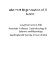 |
Aberrant Regeneration of Third Nerve | Gregory P. Van Stavern, MD | 48 year old woman S/P rupture and repair of right sided posterior communicating artery aneurysm Video shows residual partial right third nerve palsy, with aberrant regeneration, causing a pseudo Von Graefe's sign (elevation of the right upper eyelid with attempted infraduction of the right eye) Se... | Aberrant Regeneration of Third Nerve; Third Nerve Palsy |